What Prokaryote and Eukaryote Cells Worksheets Should Cover
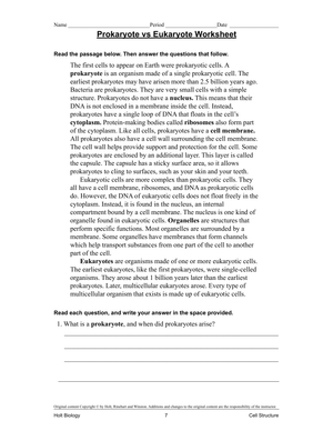
When you search for prokaryote and eukaryote cells worksheets, you're usually building a cell biology unit that needs more than a single labeling page. Teachers across grades 6 through 10 need practice materials that push students past memorizing organelle names and toward real structure-function reasoning. The strongest worksheets ask students to compare, sort, and justify their answers, not just fill in blanks and move on.
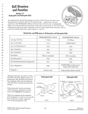
A useful set covers four things at once: the presence or absence of a membrane-bound nucleus, the organelles each cell type carries, relative size, and the cell wall differences between bacterial cells and plant cells. When those four threads run through every worksheet in the unit, students start to see why the prokaryote-eukaryote split actually matters instead of treating it as one more vocabulary list to survive.
The One Difference Students Miss Most
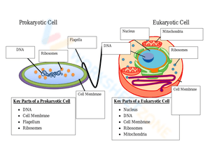
Most students can recite that prokaryotes lack a nucleus within a day of instruction. The harder idea, and the one worksheets should target, is what the two cell types share. Both prokaryotic and eukaryotic cells carry ribosomes, a cell membrane, and cytoplasm, and that overlap is exactly where sorting activities tend to fall apart. Students who treat every structure as unique to one side end up building a mental model that fails the moment a question asks about common features.
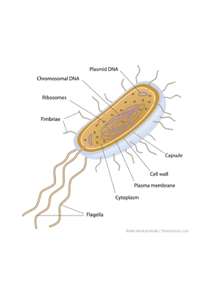
Watch for the 'ribosomes only in eukaryotes' error on any sorting task. Students reason that because ribosomes build proteins and eukaryotic cells look more complex, ribosomes must belong only to them. A quick-check item that drops a labeled ribosome into a bacterial diagram and asks true or false surfaces this misconception in under a minute, long before it shows up on a unit test. Bacteria run on ribosomes too, and that shared protein-building machinery is precisely what several antibiotics are designed to target.
Building that shared-structure check into a worksheet does more than catch one error. It teaches students that biological categories are defined by specific features, not by a vague sense of complexity. Once they internalize that ribosomes and a cell membrane show up on both sides of the sort, the rest of the comparison, from the nucleus to the cell wall, falls into place with far less reteaching.
Matching Worksheets to NGSS Standards
Cell structure sits inside a clear standards progression, and aligning your worksheets to it keeps instruction vertically coherent from one grade band to the next. NGSS MS-LS1-1 and MS-LS1-2 anchor cell structure and organelle function in grades 6 through 8, while HS-LS1-2 carries the same structure-function logic into high school biology with a deeper cell-type comparison. Building both bands into your worksheet bank lets you scaffold the same core idea year over year rather than reteaching it cold.
According to CK-12's middle school life science materials, the defining split is the nucleus: eukaryotic cells hold a membrane-bound nucleus and organelles, while prokaryotic cells do not. Prokaryotes typically measure 0.1 to 5 micrometers against the eukaryote range of 10 to 100 micrometers, a scale gap worth building directly into sorting and estimation tasks.
Naming the standard on the worksheet header helps more than it seems. When a substitute, an instructional coach, or a parent looks at the page, the MS-LS1 or HS-LS1 tag makes the purpose obvious and connects the day's practice to the year's arc. It also makes your own planning faster when you pull the same worksheet back out next fall.
Worksheet Formats That Build Structure-Function Thinking
Format drives thinking, so vary how students meet the same content. A few formats earn their place in almost every cell unit:
- Comparison charts that line up nucleus, organelles, size, and cell wall type across both cell categories, ready to use as a warm-up or an exit ticket.
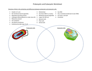
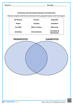
- Venn diagrams for sorting shared and unique features, which target the shared-ribosome misconception head-on.
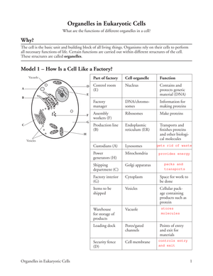
- Diagram labeling where students identify structures on unlabeled bacterial and animal cell images from memory.
- Scale problems that use the 0.1 to 5 versus 10 to 100 micrometer range so students reason about size, not just about parts.
Rotating formats keeps practice from going stale and hands you several distinct angles for formative assessment across a single week of instruction. Students who only ever label diagrams never have to defend a comparison, and that missing step is where deeper understanding usually lives.
Classroom Implementation
Sequence matters more than volume here. Open the unit with a comparison chart while students still have the vocabulary in front of them, then move to Venn sorting once they can name structures without prompts. Save diagram labeling for the point where students should be recalling from memory rather than copying. That ordering turns a stack of worksheets into a deliberate progression.
Pair the paper practice with something students can actually see. A microscope station with prepared bacterial and plant slides, or a virtual cell simulation, gives the worksheet terms a concrete anchor. Students who label a chloroplast on a page and then find one under magnification hold the prokaryote-eukaryote distinction far longer than students who only ever worked on paper.
Use short worksheets as daily entry tickets across the unit instead of one long packet. A three-item warm-up each morning tells you within minutes whether the class is ready to move forward, and it spreads the cognitive load across days instead of front-loading everything into one exhausting sitting.
Frequently Asked Questions
1. How do these worksheets help fix common cell structure misconceptions?
Sorting and Venn tasks force students to place shared structures like ribosomes and the cell membrane in the overlap, which directly confronts the belief that complex organelles belong only to eukaryotic cells. A quick true-or-false check that puts a ribosome in a bacterial diagram catches the error early, before it hardens into a habit.
2. What grade levels and standards do these worksheets fit?
They fit grades 6 through 10. NGSS MS-LS1-1 and MS-LS1-2 cover cell structure and organelle function in middle school, and HS-LS1-2 extends the comparison into high school biology, so a single worksheet bank can serve both grade bands with light adjustment.
3. Where should these worksheets sit in a cell biology unit?
Introduce them right after basic cell theory and before any organelle deep-dive. Comparison charts work as the entry point, Venn sorting fits the middle of the unit, and diagram labeling works best as review near the end, once students can recall structures without prompts.
4. How can I differentiate this content for mixed-ability classes?
Offer word banks and reduced-item charts for students who need support, and open-ended reasoning prompts about cell size and function for enrichment. The underlying content stays the same; only the cognitive demand shifts, which keeps the whole class working on prokaryote-eukaryote reasoning at once.