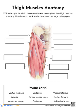
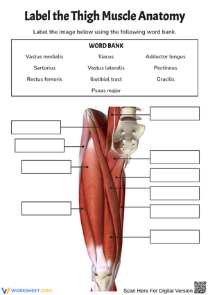
These thigh muscle worksheets pdf resources give anatomy teachers a compartment-organized approach to one of the most label-heavy units in high school life science — fifteen-plus muscles with overlapping functions, similarly constructed names, and spatial relationships that don't resolve in a standard textbook illustration. Each worksheet isolates the anterior, medial, or posterior compartment so students build accurate mental maps before encountering the full structure in a cumulative assessment.
The Three Compartments and What Students Practice
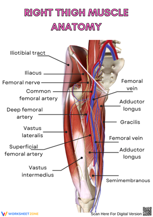
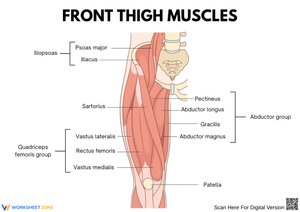
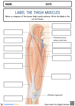
Anterior compartment worksheets center on the quadriceps femoris group and the sartorius. Students label each head of the quadriceps — rectus femoris, vastus lateralis, vastus medialis, vastus intermedius — and answer targeted questions about which head crosses the hip joint and why that distinction changes its predicted function. The sartorius gets dedicated attention because students consistently confuse its long diagonal path with the gracilis, which runs vertically through the medial compartment and looks superficially similar in most textbook illustrations.
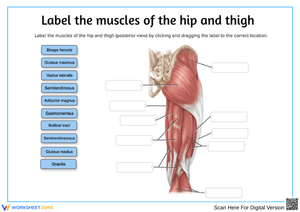
Posterior compartment work focuses on the hamstrings: biceps femoris, semitendinosus, and semimembranosus. Students trace each muscle from the ischial tuberosity to its distal attachment, then mark which muscles terminate on the medial versus lateral side of the knee. Clinical injury questions in this section ask students to explain why two-joint muscles are especially vulnerable during explosive movements — a direct application of origin-insertion knowledge that students interested in athletic training find immediately relevant.
Medial compartment exercises address the full adductor group: pectineus, adductor brevis, adductor longus, adductor magnus, and gracilis. Students sort these muscles by depth and by distal attachment site — tibial versus femoral — to build precision beyond name recall. The gracilis worksheet draws an explicit comparison to the sartorius, since both are long strap muscles crossing two joints, a pairing that reinforces the anterior-medial boundary rather than blurring it. A well-constructed thigh muscle worksheets pdf includes cross-sectional views alongside surface views specifically to help students locate muscles like the adductor brevis, which simply cannot be traced on a standard anterior diagram.
Predictable Errors to Address Before the Assessment
The most reliable error in the anterior compartment is omitting or mislabeling the vastus intermedius. It sits deep to the rectus femoris and disappears entirely in surface-view illustrations, so students who have only seen standard diagrams skip it or write its name next to the vastus medialis. A worksheet that pairs a surface view with a mid-thigh cross-section eliminates that avoidance strategy — there is no way to label the cross-section correctly without locating it independently of the overlying rectus femoris.
In the medial compartment, name confusion is near-universal and follows a specific pattern. Students who correctly identify adductor longus on a Monday exercise write "adductor magnus" in that spot by Friday because the names are similar enough to drift without a functional anchor. Tasks that require students to rank the adductors from most superficial to deepest, or to identify which one has a tibial rather than a femoral attachment, give those names something spatial and functional to cling to rather than leaving them as a list to memorize and forget.
The hamstring section produces a subtler but consistent error: students write that all three hamstrings attach to the tibia, missing the biceps femoris's insertion on the fibular head. In actual student work this surfaces on the lateral-versus-medial sorting task — the student has absorbed a general rule ("hamstrings attach below the knee") without examining the specific anatomy of each muscle individually.
Working These Worksheets Into Your Unit Plan
Most anatomy teachers who use this set open each compartment with a whole-class diagram on the whiteboard, then move students to the corresponding worksheet for the last fifteen minutes of the period. That worksheet doubles as the evening review assignment, and the following morning's warm-up is a short matching check drawn directly from the same material. That three-day rhythm — introduce, practice, retrieve — benefits thigh anatomy specifically because the volume of muscle names rewards spaced retrieval far more than a single intensive exposure the night before an exam.
The clinical case worksheets fit best after students have completed all three compartment diagrams. A scenario like "a soccer player feels a sudden pop at the back of the thigh during a sprint" requires students to pull simultaneously from their posterior compartment diagram and their understanding of two-joint muscle vulnerability. Used that way, the case study functions as an integration task rather than a standalone reading exercise, and it naturalizes the connection between anatomical structure and real injury mechanisms before the unit ends.
A thigh muscle worksheets pdf from this set also serves as a formative check mid-unit. The origin-insertion matching page takes about eight minutes at the end of a class period, tells the teacher immediately which compartment needs re-teaching, and gives students a low-stakes chance to surface their own gaps before the summative assessment.
Standard Alignment
These worksheets align directly to NGSS HS-LS1-2 (Structure and Function), which requires students to construct explanations for how the structural organization of interacting body systems produces coordinated movement. The labeling and function-prediction tasks address that standard precisely — students are not simply identifying muscles by name but explaining how attachment points determine direction of pull, which is the structural-to-functional reasoning HS-LS1-2 demands. Within a typical anatomy unit, this standard is first addressed at the cellular and tissue level before scaling to organ systems; the thigh is a productive anchor at the muscle level because it presents all three functional directions — flexion, extension, and adduction — within one anatomical region, making the structure-function relationship visible without requiring students to jump between body regions mid-unit.
Differentiating the Set for Mixed-Ability Classrooms
For students who find the full compartment diagram overwhelming, break the anterior worksheet into two passes: label only the four quadriceps heads first, confirm accuracy, then return to add the sartorius. Reducing the number of simultaneous demands on working memory without changing the content standard keeps the task manageable without watering down the anatomy.
Students who move through the material quickly find the clinical case worksheets genuinely demanding because those exercises require synthesis rather than recall. Asking those students to write a full mechanism-of-injury explanation — naming the specific muscles involved, identifying the stressed attachments, and explaining why two-joint architecture increases vulnerability — produces analytical responses that go well beyond what a standard labeling task can assess. A thigh muscle worksheets pdf set that includes both a basic labeling tier and a clinical application tier serves a mixed-ability class without requiring two entirely separate lesson plans.
Frequently Asked Questions
Which muscles make up the quadriceps femoris, and do all four act on the same joints?
The quadriceps group consists of four heads: the rectus femoris, vastus lateralis, vastus medialis, and vastus intermedius. All four converge on the patella via the quadriceps tendon and continue to the tibial tuberosity, functioning together as the primary extensors of the knee. However, only the rectus femoris originates on the anterior inferior iliac spine rather than the femoral shaft, which means it crosses the hip joint and assists with hip flexion — a distinction the other three heads do not share. This is the functional detail that separates routine recall from actual anatomical understanding on any unit assessment.
Why are the hamstrings described as two-joint muscles, and why does it matter clinically?
The hamstrings — biceps femoris, semitendinosus, and semimembranosus — all originate on the ischial tuberosity and cross both the hip and knee joints, extending the hip and flexing the knee simultaneously. Because the muscle must accommodate position changes at two joints at once during explosive movements, any sudden shift that stretches one end while the other contracts generates exceptional tensile stress. Hamstring strains are among the most common soft-tissue injuries in sprinting sports precisely because of this two-joint architecture, which is why the clinical case worksheets return to origin-insertion relationships rather than asking students only to name the muscles.
Students keep mixing up the sartorius and the gracilis — is there a reliable way to teach the distinction?
The visual anchor that works most reliably is path direction. The sartorius runs diagonally across the anterior thigh from the anterior superior iliac spine down to the medial proximal tibia — a line that cuts across the compartment. The gracilis runs nearly vertically along the medial thigh from the pubis to the same tibial attachment. Having students draw and physically trace those two lines on a blank thigh outline before labeling them tends to fix the distinction in a way that reading definitions does not. The sartorius also belongs to the anterior compartment based on innervation by the femoral nerve, despite its medial distal attachment — a fact worth stating explicitly when students ask why it isn't grouped with the adductors.
Is there a reliable strategy for keeping the adductor group names sorted?
The mnemonic Pigs Always Bring Large Muddy Grins maps to pectineus, adductor brevis, adductor longus, adductor magnus, and gracilis — moving roughly superficial to deep and superior to inferior. That said, a mnemonic only holds when it attaches to something spatial, so pair it with a depth-ranking task on the medial compartment worksheet rather than treating it as a standalone memory device. Students who can arrange the muscles in order on a diagram, not just recite the mnemonic in sequence, retain the information past the exam date.