Calf Muscle Printable Worksheets for Anatomy Class
These calf muscle printable worksheets give anatomy and life science teachers a ready-made structure for turning a muscle students have already felt — during every sprint, stair climb, or nighttime cramp — into a formal case study in origin and insertion, fiber type, and joint mechanics. Six worksheet formats span from vocabulary identification to injury-based application, covering both the gastrocnemius and the soleus in enough anatomical depth for a high-school A&P course while remaining workable in a middle-school life science unit.
The Anatomy Covered Across the Set
The posterior compartment of the lower leg gives students a compact, manageable region: two primary muscles, one shared tendon, one innervating nerve, and one fundamental joint action. That tightness is pedagogically useful — students can genuinely master this region rather than skim twelve muscles and retain none of them.
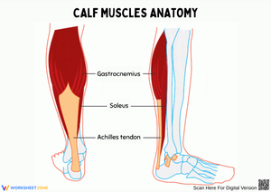
The gastrocnemius is the muscle students can see and palpate on their own leg. Its medial and lateral heads originate on the femoral condyles, positioning it above the knee and making it one of the few lower-leg muscles that crosses two joints. That bi-articular characteristic generates a genuine conceptual shift: students who have assumed every muscle acts at the joint closest to it must revise that assumption here. The muscle's dominance in fast-twitch fibers explains its role in explosive push-off during jumping and sprinting, and that fiber-type concept connects naturally to exercise physiology discussions later in the course.
The soleus lies beneath the gastrocnemius and crosses only the ankle joint, restricting its action to plantarflexion. Its slow-twitch fiber composition suits sustained work — standing, walking uphill, long-distance running — and its role as a venous pump gives teachers a built-in bridge to the circulatory system. Both muscles converge into the Achilles tendon, the largest tendon in the body, which inserts on the calcaneus. The tibial nerve, arising from spinal levels S1–S2, innervates both muscles — a detail that connects this unit to nervous-system content and helps students understand why certain nerve compressions weaken plantarflexion before any formal neuroscience instruction arrives.
Six Worksheet Formats, Each With a Distinct Purpose
The calf muscle printable worksheets in this set are deliberately varied in task type because repeating the same format — label, label, label — produces diminishing returns by the second session. Each worksheet asks students to process the same anatomical content through a different cognitive lens.
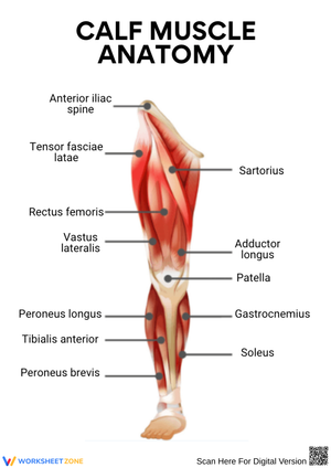
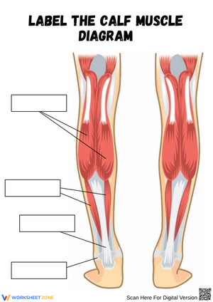
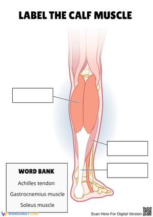
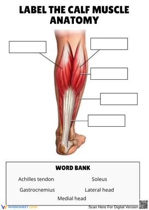
- Label-the-Diagram: A blank illustration of the posterior lower leg. Students label the gastrocnemius (both heads), soleus, Achilles tendon, calcaneus, tibia, and fibula. Builds spatial awareness and anatomical vocabulary simultaneously.
- Compare-and-Contrast Chart: Two columns — gastrocnemius versus soleus — across categories including location, joints crossed, predominant fiber type, primary action, and a clinical note. Pushes students from identification toward analysis.
- Fill-in-the-Blank Passage: A short reading with key terms removed — plantarflexion, Achilles tendon, tibial nerve, calcaneus — so students practice retrieval in context rather than in isolation.
- Function-Matching Activity: Students draw lines or write letters to match each muscle to its action, joints crossed, origin, insertion, and nerve supply. Works efficiently as a formative check before a quiz.
- Cross-Section Diagram: A transverse view of the lower leg asks students to locate the posterior compartment and distinguish the gastrocnemius layer from the soleus layer beneath it. Reading cross-sectional anatomy is a distinct visual skill — one that matters for any future work involving imaging or lab dissection.
- Injury-Connection Worksheet: Students read three brief scenarios — a calf strain during a basketball game, Achilles tendinitis in a distance runner, and nighttime leg cramps — then identify the affected structure and explain the anatomical reasoning. This format resonates with student athletes, who bring prior experience to the analysis.
The Errors That Surface Consistently in This Unit
The most persistent mistake is collapsing the gastrocnemius and soleus into a single structure. On the label-the-diagram worksheet, students routinely write "gastrocnemius" across the entire posterior calf without placing the soleus beneath it. The cross-section worksheet addresses this directly: a transverse view forces students to think in anatomical layers rather than surface silhouettes, and the error nearly always resolves after one session with that format. The cross-section worksheet is also the one that benefits most from a brief teacher demonstration before students work independently — students who have never read a transverse anatomical view will orient incorrectly without a 90-second explanation of the viewing angle.
Origin-and-insertion reversal is the second reliable error. Students can recite the definitions — origin is the fixed attachment, insertion is on the moving bone — but under pressure they assign them backwards, writing that the gastrocnemius inserts on the femoral condyles. The function-matching worksheet surfaces this quickly because both attachment points appear in the same task. Having students underline "fixed" and "moving" in the definitions before starting reduces the reversal rate noticeably in practice.
Fiber type is the hardest concept to transfer from worksheet to quiz. Students can repeat "fast-twitch equals explosive power, slow-twitch equals endurance" as isolated facts, then write "fast-twitch" for the soleus because they associate the calf in general with running speed. The compare-and-contrast chart places the fiber-type row directly beside the primary-action row, making the relationship between composition and function visible side by side rather than implicit in memory. Students who annotate the chart with their own explanatory notes — even a single sentence — retain the distinction at higher rates than those who fill in the boxes and move on.
Sequencing These Worksheets Across a Three-Day Mini-Unit
A three-day sequence works well for this set. Day one uses the label-the-diagram and fill-in-the-blank worksheets to establish the vocabulary students need before any comparative analysis is possible. Day two introduces the compare-and-contrast chart and the function-matching activity, shifting the cognitive demand from recall to comparison — students who built the vocabulary on day one can now use it to draw distinctions rather than just reproduce terms. Day three deploys the injury-connection worksheet as an application task and closes with the cross-section diagram as a formative check on spatial understanding. That three-step movement from knowledge to analysis to application requires no additional lecture material between worksheet sessions.
For pairing with kinesthetic activity: have students perform ten calf raises at their desks, then immediately annotate the label-the-diagram worksheet to indicate which muscle is doing the primary work. The sensation is still present, and the annotation task gives it anatomical language. If resistance bands are available, students plantarflex against load and complete the function-matching worksheet while the proprioceptive experience is fresh. Station rotations work well too — one station with an anatomy model or poster, one with a worksheet, one with a short projected video clip — rotating every eight to ten minutes. The calf muscle printable worksheets fit station formats cleanly because each one is self-contained and requires no verbal setup to begin.
Standard Alignment
At the middle-school level, this content connects to NGSS MS-LS1-3, which asks students to explain how body systems interact to support life functions — the soleus's venous pump role and the calf's contribution to locomotion are direct examples. At the high-school level, NGSS HS-LS1-2 addresses how the structure of biological systems determines their function; the gastrocnemius-soleus fiber-type comparison illustrates that principle at the tissue level in a way students can observe on their own bodies. For courses aligned to the National Health Science Standards, particularly Standard 1 (Academic Foundation), these worksheets address anatomical structure and physiological function within the musculoskeletal system, with the injury-connection worksheet extending into pathology awareness and applied health literacy.
Making the Set Work Across Different Readiness Levels
The label-the-diagram worksheet adjusts easily across readiness levels. Students who need additional support receive the diagram with a word bank; on-level students receive the same diagram without it. For advanced students, adding the plantaris and popliteus to the structure list creates a genuine extension: the plantaris has a notably long, thin tendon sometimes harvested in reconstructive surgery — a detail that functions as a research hook rather than arbitrary extra content.
The compare-and-contrast chart extends to a third column for the plantaris as a challenge task. The fill-in-the-blank passage works in two printed versions: one with partial cues — first letters of each missing term — for students who need a lighter load, and one with no cues for on-level and above-level students. For the injury-connection worksheet, students who need more support work with a reference card listing each muscle's location, joints crossed, and primary action; advanced students add a brief treatment or rehabilitation rationale that requires applying anatomical knowledge beyond simply identifying the affected structure. These calf muscle printable worksheets are formatted so those adjustments take a few minutes to configure before printing, not a full redesign of the activity.
Frequently Asked Questions
Are these worksheets appropriate for both middle school and high school?
Yes. The anatomical content — gastrocnemius, soleus, Achilles tendon, tibial nerve, plantarflexion — appears in both middle-school life science and high-school anatomy and physiology curricula. The label-the-diagram and fill-in-the-blank worksheets work at the middle-school level, where vocabulary building is the primary goal. The compare-and-contrast chart, function-matching activity, and injury-connection worksheet carry more analytical weight and are better suited to high school, though advanced middle-school students handle them without difficulty.
How do the gastrocnemius and soleus differ in function?
Both muscles plantarflex the ankle, but the gastrocnemius also assists knee flexion because it crosses the knee joint. The soleus crosses only the ankle and contributes exclusively to plantarflexion. Their fiber composition differs as well: the gastrocnemius is fast-twitch dominant and produces explosive force, while the soleus is slow-twitch dominant and sustains postural and endurance work. These distinctions are the central focus of the compare-and-contrast chart and the function-matching worksheet.
Which nerve innervates the calf muscles, and why teach that detail?
The tibial nerve, arising from spinal levels S1–S2, supplies both the gastrocnemius and the soleus. Including innervation in the unit helps students understand why a herniated disc at the L5–S1 level — or a direct tibial nerve injury — can produce weakness in plantarflexion. That clinical connection makes the nervous-system unit feel relevant well before students formally reach it in the course sequence.
Can these worksheets serve as graded assessments rather than practice?
Several adapt readily. The label-the-diagram worksheet without a word bank tests pure recall under timed conditions. The compare-and-contrast chart becomes a rubric-scored performance task when students write complete sentences explaining each difference rather than filling in single-word boxes. The injury-connection worksheet functions as a higher-order thinking assessment — open-note or closed — because it asks students to apply anatomical knowledge to scenarios they have not seen before, rather than reproduce memorized definitions.
Clear All