These lower back muscles worksheets pdf resources give anatomy teachers a set of ready-to-print labeling, matching, and clinical application exercises that make the lumbar region genuinely comprehensible to high school students — not just a list of Latin terms to survive a test. Each worksheet targets a distinct skill, so teachers can distribute them across a unit rather than front-loading everything into a single class period. The set is built around the three muscle groups students encounter most consistently in a high school Anatomy & Physiology course: the erector spinae, the latissimus dorsi, and the quadratus lumborum.
Muscles and Tasks in Each Worksheet
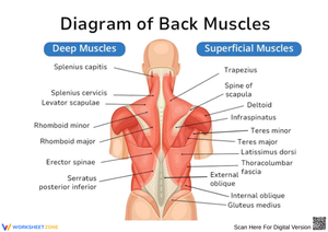
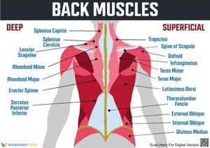
The labeling worksheets use posterior-view diagrams at two levels of anatomical depth. The first presents only the superficial layer — the latissimus dorsi and the visible portions of the erector spinae — so students establish a baseline before confronting deeper musculature. The second diagram adds the quadratus lumborum and asks students to annotate each structure with its primary action, not just its name. Students who can recite muscle names but cannot explain what those muscles do during movement show up immediately on that second worksheet.
A function-matching exercise pairs each muscle with a specific movement: spinal extension, lateral flexion, pelvic elevation during single-leg stance. A clinical application worksheet rounds out the set with brief case descriptions — a cross-country runner presenting with unilateral lumbar tightness, a student who sits for six hours in a school day — and asks students to identify which muscles are likely involved, what movements would be painful, and what the anatomy suggests about recovery. That kind of problem ties anatomical identification to physiology and health literacy, which is where the content becomes memorable for most teenagers.
Frequent Student Errors Worth Anticipating
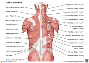
The most reliable mistake in lumbar anatomy is treating the erector spinae as a single structure. Students who have correctly learned that it comprises iliocostalis, longissimus, and spinalis will still write "erector spinae" across every blank on a diagram that specifically calls for component names. The solution is a worksheet that forces column-by-column identification — not a holistic label with room to be vague.
The quadratus lumborum causes a different problem. Most students reduce it to "lateral flexion" and stop there, because that is the action they encounter first. They consistently miss its stabilizing role during contralateral leg lift — the reason a QL strain makes walking painful before it makes forward bending painful. Worksheets that include functional scenarios rather than movement lists alone surface this gap directly, without requiring additional teacher intervention to uncover it.
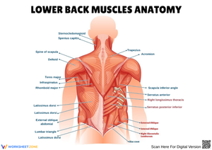
The latissimus dorsi surprises students who have only seen it labeled in upper-body diagrams. Because it features prominently in pull-up illustrations, students frequently place its origin in the mid-thoracic region and ignore its broad attachment along the lumbar fascia and posterior iliac crest. A posterior-view diagram that extends from the thoracic vertebrae to the sacrum corrects this without a separate lecture.
How to Build These Worksheets Into Your Lesson Plan
The superficial-layer labeling diagram works well as an entry task on the first day of spinal musculature instruction. Students attempt the posterior view using whatever prior knowledge they bring — the gaps in their initial attempts tell you precisely where direct instruction needs to focus. During the core lesson, the function-matching exercise runs as guided practice: as each muscle's action is explained, students fill in the corresponding row. Having students stand briefly and perform a controlled spinal extension while looking at the erector spinae label is worth the ninety seconds it takes. The motor memory anchors the anatomy in a way that passive note-taking does not.
The clinical application worksheet belongs at the end of the sequence, either as homework after the lumbar unit closes or as a short formative check at the start of the following class. Students who genuinely understand the content complete it quickly; students who memorized without understanding stall on the second case. That contrast identifies exactly who needs another pass at the material before any summative grade is recorded, which makes the worksheet worth assigning even when it won't appear directly on a test.
Standard Alignment
A lower back muscles worksheets pdf aligns most directly with NGSS standard HS-LS1-2, which asks students to "develop and use a model to illustrate the hierarchical organization of interacting systems that provide specific functions within multicellular organisms." The lumbar region is an unusually clean teaching site for this standard because the muscle layers are genuinely hierarchical — superficial, intermediate, and deep — and the functional interdependence between the muscular and skeletal systems is visible in every movement the lower back performs. When a student diagrams how the erector spinae and quadratus lumborum cooperate to stabilize the spine during lateral flexion, they are doing the systems-level thinking this standard targets. The labeling and annotation tasks also generate written artifacts that document that thinking, which is useful when standards documentation is required.
Differentiating These Worksheets for a Mixed-Ability Classroom
For students who struggle with anatomy vocabulary, the labeling worksheets can run with a word bank that lists all muscle names. This removes the retrieval barrier and lets students focus on spatial placement — the actual skill being built at that stage. The function-matching exercise can be modified similarly: provide the movement column pre-filled and ask students only to identify the matching muscle, rather than generating both columns from memory.
Advanced students or those in an AP-level course benefit most from the clinical application worksheet extended with a full anatomical explanation requirement: not just "the QL is involved" but "the left quadratus lumborum is likely strained because pelvic elevation during right-leg stance requires eccentric control of the contralateral side." In mixed-ability sections, teachers can assign the labeling task universally and reserve that extended clinical reasoning as an enrichment layer for students who finish early, without creating a separate assignment entirely.
Frequently Asked Questions
At what point in an anatomy unit do these worksheets fit best?
The labeling and function-matching worksheets belong in the middle of a muscular system unit, after students have covered basic muscle tissue structure and before the class moves into the upper extremity. The clinical application worksheet works best at the end of the lumbar sequence as a bridge into health applications or injury analysis. A lower back muscles worksheets pdf is not a standalone unit — it functions as one focused component inside a larger sequence on axial musculature, and it works best when teachers have already introduced origins, insertions, and actions as a general framework.
Do the worksheets include answer keys?
Yes. Each worksheet comes with a fully labeled answer diagram and a completed function-matching key. The clinical application worksheet includes sample responses that identify the relevant muscles, the expected movement impairments, and the anatomical reasoning behind each case. Those keys make it practical to assign the worksheets as independent practice or homework without turning every round of grading into a time-intensive project.
How do the diagrams handle the layering problem in lumbar anatomy?
The set uses two separate posterior-view diagrams — one showing the superficial layer and one revealing the deeper stabilizing muscles — rather than a single image that overlaps everything at once. Presenting layered anatomy on one crowded diagram forces students to manage visual complexity and content complexity simultaneously, which is more than most students can handle in a single pass. Separating the layers into two worksheets keeps cognitive load manageable and gives teachers a natural two-day sequence: superficial muscles on day one, deep stabilizers on day two. A lower back muscles worksheets pdf that handles layering this way produces noticeably cleaner student work and far fewer blank answers at the deep-muscle level than single-diagram formats do.
Are these worksheets appropriate for a general biology course, or only Anatomy & Physiology?
The labeling worksheets work in either context. A general biology course covering body systems can use the superficial-layer diagram without requiring the clinical depth that an A&P course demands. The function-matching and clinical application worksheets are better suited to A&P, where students have the prerequisite understanding of muscle physiology — origin, insertion, action — that those exercises require. Teachers in a general biology course should preview the clinical application worksheet before assigning it; students without a grounding in muscle terminology will find the case descriptions harder to parse than the diagram work, and the frustration is not productive at that level.