These pectoral muscle worksheets printable materials give science teachers a structured path through one of the most spatially demanding topics in a muscular system unit — the chest region, where two muscles occupy different anatomical planes and students must track both simultaneously. Each worksheet targets a specific layer of that challenge, from basic identification to origin-insertion mapping and functional movement analysis.
What Students Work Through
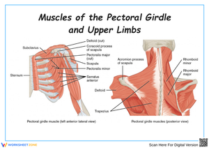
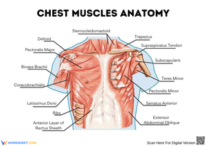
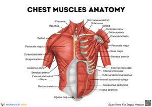
The pectoralis major and pectoralis minor look manageable on a diagram until students attempt to label them from memory. The major originates across two distinct heads — clavicular and sternocostal — that function as one muscle with multiple anchor points. The minor adds a depth relationship that flat diagrams rarely communicate clearly. Each worksheet addresses one or two of these concepts at a time rather than requiring students to hold the full picture at once.
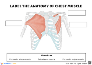
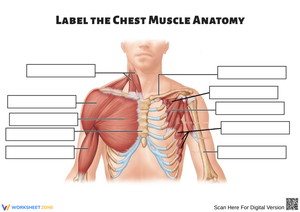
- Labeling both muscles on anterior and lateral thorax views
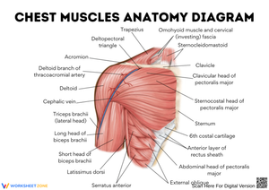
- Identifying the clavicular and sternocostal heads of the pectoralis major and tracing their convergence toward the intertubercular groove of the humerus
- Locating the rib 3–5 attachment points for the pectoralis minor and its insertion at the coracoid process of the scapula
- Drawing directional arrows for adduction and internal rotation at the glenohumeral joint
- Mapping the depth relationship between both muscles on layered anterior-view diagrams
- Annotating the medial and lateral pectoral nerve pathways for upper-level work
Frequent Student Errors Worth Watching For and Correcting
The most consistent error is origin-insertion reversal on the pectoralis major. A student accurately sketches the muscle's fan-shaped belly and correctly identifies the humerus as an attachment site — then labels it the origin, because the humerus is visually prominent in the diagram. The clavicular and sternal attachments, which form the actual origin, read to many students as destinations rather than starting points. Worksheets that ask students to draw the line of pull, not just label two points, expose this reversal immediately.
A second predictable error involves the pectoralis minor's insertion. Students who recently studied the deltoid frequently point to the acromion instead of the coracoid process — both sit on the scapula, both serve as muscle attachment references in the same unit, and the acromion is simply more familiar. Worksheets presenting the scapula with unlabeled bony landmarks force students to locate the coracoid process independently rather than pattern-match to a labeled key.
A subtler gap shows up in how students interpret depth. Most understand the verbal statement that the pectoralis minor lies deep to the major, but their diagrams place both muscles at the same visual plane. This is a spatial reasoning problem, not a vocabulary problem. Layered worksheets — presenting the minor first, then overlaying the major in a second view — make the three-dimensional relationship visible where a single composite diagram leaves it abstract.
Working These Worksheets Into Your Weekly Schedule
The chest muscles typically land in the second or third week of a muscular system sequence — after students can read a diagram independently but before origin-insertion logic has become automatic. That timing is where these worksheets carry the most instructional weight.
Before any diagram comes out, run a brief kinesthetic moment: have students press their palms together in front of their chest in an isometric hold — a direct contraction of the pectoralis major — and ask them to notice where the tension sits. The front of the chest wall, the sternum, the anterior shoulder. When the worksheet follows immediately, students trace the muscle's origin across those same regions with physical memory backing the visual recognition. That two-minute activity measurably reduces the blank-stare response on the subsequent labeling task.
The five-to-eight minute block before dismissal or after a content-heavy lecture works well for one targeted worksheet. The format asks for a bounded task — label these structures, draw this line of pull — rather than open-ended note-taking, which collapses when students are already cognitively overloaded. These pectoral muscle worksheets printable activities also function as self-contained substitute lesson plans; the diagram context is sufficient for students to begin without live teacher explanation.
Standard Alignment
These worksheets align to NGSS HS-LS1-2, which asks students to develop and use a model to illustrate the hierarchical organization of interacting systems that provide specific functions within multicellular organisms. In classroom terms, that standard appears when students draw and annotate the relationship between skeletal attachment points, the muscle belly, and the resulting joint movement — exactly what the origin-insertion and movement-arrow exercises require. State anatomy and physiology standards covering musculoskeletal structure and function in the upper limb map directly onto the same content.
Adapting the Set for a Range of Learners
For students still building anatomical vocabulary, the pre-labeled diagrams serve as study guides. A quick protocol — underline every term you can define, circle every term you need to review — takes ninety seconds and surfaces the exact gaps before a quiz. That self-assessment habit is worth establishing early in the unit, well before summative assessment pressure arrives.
Students past surface identification use the blank skeletal torso format: draw both muscles from scratch, indicate lines of pull, write a two-sentence functional explanation beneath each. For advanced anatomy students, the innervation exercises — tracing the medial and lateral pectoral nerves from the brachial plexus to their respective muscles — extend the same diagram into genuinely demanding territory. These pectoral muscle worksheets printable resources cover that full range without requiring separate materials for each tier of the class.
Frequently Asked Questions
What distinguishes the pectoralis major from the pectoralis minor on these worksheets?
The worksheets present both muscles in anatomical context: the pectoralis major as the large, fan-shaped superficial muscle visible on the anterior chest, and the pectoralis minor as the smaller, triangular muscle lying entirely beneath it. Several worksheets require students to draw the depth relationship explicitly — placing the minor in a deep view before adding the major on top — rather than simply identifying two labeled structures on the same diagram.
Are these worksheets suited to both introductory and advanced anatomy courses?
The set moves from basic identification through origin-insertion mapping to nerve pathway annotation. An introductory class works with the labeled diagrams and coloring activities; an advanced class uses the blank skeletal outlines and innervation exercises. The depth of the task sets the difficulty level — teachers do not need to rewrite or modify the core diagrams to use them at different levels.
How do blank diagrams work without students shutting down when they feel lost?
Run the kinesthetic activity first so students enter the blank diagram with a felt physical reference rather than a blank mental model. Then allow three to four minutes of independent work before a brief peer-check — partners compare their labeling before the teacher reveals the correct version. That sequence produces more durable retention than handing out a filled-in answer key and generates enough visible misconceptions that whole-class corrections feel targeted rather than generic.
What grade levels and courses are these written for?
The content suits high school anatomy and physiology courses, typically grades 10–12. Biology courses that include a muscular system unit draw on the introductory worksheets. AP Biology and dedicated anatomy electives use the full set, including the advanced innervation activities. These pectoral muscle worksheets printable materials are not written for middle school science, where the muscular system is taught at the organ-system level without the specificity of individual muscle origin and insertion points.