These foot muscle worksheets pdf resources give anatomy teachers something that broad lower-limb units rarely deliver: sustained, focused practice on one of the body's most structurally dense regions. The human foot contains more than 20 intrinsic muscles arranged across four plantar layers, plus a set of extrinsic muscles originating in the lower leg—and students in AP Biology, college Anatomy and Physiology, and allied health programs are expected to name them, locate them, and describe their actions before a unit exam.
Anatomy Content Across the Set
Each worksheet targets a specific layer of foot muscle knowledge, building from spatial orientation to functional recall. The set addresses both intrinsic muscles—those that originate and insert entirely within the foot—and extrinsic muscles, which arise in the lower leg and act on the foot through long tendons that cross the ankle joint.
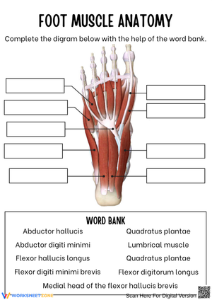
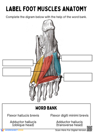
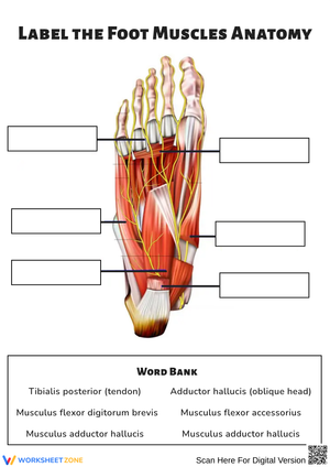
- Plantar layer labeling: Blank diagrams cover all four plantar layers in sequence—the superficial layer containing the flexor digitorum brevis and abductor hallucis, the second layer with the quadratus plantae and lumbricals, the third layer housing the flexor hallucis brevis and adductor hallucis, and the deep fourth layer with the interossei.
- Dorsal surface identification: A separate worksheet focuses on the extensor digitorum brevis and extensor hallucis brevis, two intrinsic muscles students frequently overlook when studying from plantar-only diagrams.
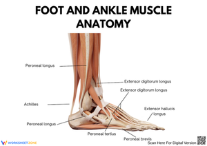
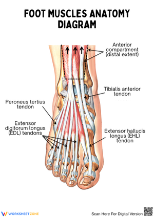
- Extrinsic muscle tracing: Students trace the path of muscles like the tibialis anterior, peroneus longus, and flexor hallucis longus from their lower-leg origins to their foot insertions, reinforcing why lower-leg injuries can produce foot-level symptoms.
- Origin-insertion-action tables: Structured tables prompt students to record each muscle's proximal attachment, distal attachment, and primary movement—a format that transfers directly to practical exams and clinical reasoning tasks.
- Function-matching exercises: Students match muscle names to actions such as toe flexion, arch support, digit abduction, and eversion, building the functional vocabulary anatomy assessments require.
Anatomy references including Teach Me Anatomy and the Kenhub muscle database organize the plantar surface into four distinct functional layers—and each foot muscle worksheets pdf in this set mirrors that layered structure so students build a spatial map rather than a memorized list of names.
Errors Students Make That These Worksheets Help You Catch
The most persistent confusion in student work is between intrinsic and extrinsic classifications. Students often assume "intrinsic" simply means "small" and will list the peroneus longus as intrinsic because it seems to do foot-level work. The misunderstanding runs deeper than vocabulary: they have not yet internalized that origin location—not muscle size or function—determines the classification. A worksheet that asks students to mark origin points on both a leg diagram and a foot diagram, before they label anything else, addresses this at the source.
A second reliable error involves the flexor hallucis longus and flexor hallucis brevis. Students who correctly identify the brevis on a plantar diagram will still write "flexor hallucis longus" in the same blank because the names are nearly identical and both muscles flex the big toe. Worksheets that require students to annotate each muscle's origin—not just its name—force the distinction in a way that a multiple-choice question cannot.
Layer confusion is the third pattern worth anticipating. When all four plantar layers appear on a single diagram, students compress the visual information and routinely misplace the adductor hallucis (layer 3) into layer 2 and the interossei (layer 4) into layer 3. Presenting each layer on its own worksheet diagram rather than stacking all four into one figure reduces this specific error substantially.
How to Work These Worksheets Into Your Anatomy Unit
The most effective sequence starts not with labeling but with orientation. On the first day of the foot unit, use a coloring worksheet—one for each plantar layer—so students encounter the muscle arrangement as a progressive reveal rather than an all-at-once inventory. Cognitive load research supports this directly: presenting all four plantar layers simultaneously before students know any muscle names pushes working memory beyond capacity. Each layer needs its own processing time before the full picture is meaningful.
By day three, blank-diagram labeling works well as a low-stakes checkpoint before students open their notes—this is exactly when the intrinsic-vs-extrinsic confusion surfaces most visibly, and catching it here leaves two class periods to address it before the formal assessment. For allied health students specifically, the foot muscle worksheets pdf format allows instructors to add clinical annotation tasks directly to a printed diagram: circling the muscles implicated in plantar fasciitis, marking the arch-support structures compromised in flatfoot presentation, or tracing the tendon path disrupted by a lateral ankle sprain.
Origin-insertion-action tables belong toward the end of the sequence, not the beginning. Students who complete them after they can already label a diagram work through the tables with existing spatial anchors—each muscle name connects to a location in memory, not just an empty row. Self-grading with answer keys on the final review day prompts the kind of error-by-error discussion that makes group review genuinely productive rather than performative.
Adjusting the Worksheets for Students at Different Levels
For introductory-level students or those newer to anatomical vocabulary, a reasonable entry point is restricting the first labeling worksheet to plantar layer one only: the flexor digitorum brevis, abductor hallucis, and abductor digiti minimi. Three muscles with distinct locations and functions give students enough to practice identifying a structure, recording its name, and describing its action without the cognitive weight of the full set. Once layer one is solid, layers two through four follow in order.
Advanced students in college A&P or pre-clinical programs benefit from an additional task layer: adding columns for nerve supply and arterial source alongside the standard origin-insertion-action data, or including a "clinical note" column where students must name one pathological condition associated with dysfunction of that muscle. That shift from identification toward applied reasoning is appropriate for students preparing for board-style exams in nursing, physical therapy, or kinesiology programs.
Standard Alignment
These worksheets align with NGSS standard HS-LS1-2, which asks students to develop and use a model to illustrate the hierarchical organization of interacting systems that provide specific functions within multicellular organisms. In classroom terms, this standard becomes most relevant when students move from naming individual muscles to understanding how those muscles operate as coordinated functional units—exactly what the plantar-layer sequence and extrinsic-tendon tracing exercises build. Instructors in college A&P courses using the HAPS Learning Outcomes Framework will find the worksheets map directly to the musculoskeletal module outcomes covering muscle identification, attachment site anatomy, and movement production.
Frequently Asked Questions
What is a realistic scope for a high school anatomy unit on foot muscles?
Most high school AP Biology courses have instructional time for the first two plantar layers in depth—the flexor digitorum brevis, abductor hallucis, quadratus plantae, and lumbricals—plus the primary extrinsic muscles: tibialis anterior, peroneus longus, and the flexor and extensor hallucis longus. Layers three and four, including the adductor hallucis and both sets of interossei, are more appropriate for college A&P and allied health courses where clinical application is built into the curriculum.
How do students learn to distinguish intrinsic from extrinsic foot muscles?
The fastest path is a diagram that shows both the lower leg and the foot simultaneously, with marked lines tracing where each muscle begins. When students see the tibialis anterior originating on the lateral tibial condyle and following its tendon path across the ankle before inserting into the medial cuneiform, the "extrinsic" label becomes self-evident rather than arbitrary. A worksheet that presents origin points visually—rather than as text in a table—makes this distinction stick in a way that reading a definition rarely does.
Are these worksheets appropriate for physical therapy or kinesiology students?
Each foot muscle worksheets pdf in this set works directly in allied health coursework. Physical therapy and kinesiology students benefit especially from the extrinsic-muscle tracing exercises and any format connecting muscle location to movement dysfunction. Instructors in those programs typically add clinical application questions to the standard labeling and table tasks—identifying which muscles are disrupted in plantar fasciitis, posterior tibial tendon dysfunction, or peroneal nerve injury—and the diagrams in this set carry enough anatomical detail to support that analysis.