These deltoid muscle worksheets give anatomy teachers six distinct activity types for one of the shoulder's most instructionally rich muscles — a structure students can name on sight but rarely analyze with real precision. Each worksheet targets a specific cognitive demand: labeling bony landmarks, matching functional heads to their actions, charting origins and insertions, reading for clinical application, diagramming movement planes, or comparing the deltoid against the rotator cuff group. The set builds layered understanding that holds through unit exams and into lab practicals on anatomical models.
The Specific Skills Each Worksheet Targets
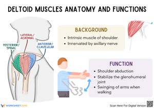
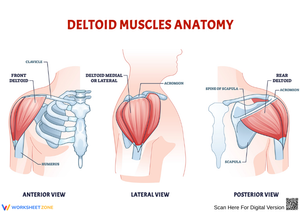
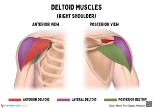
The deltoid's three heads — anterior (clavicular), middle (acromial), and posterior (spinal) — share a single insertion at the deltoid tuberosity of the humerus but perform entirely different movements. The anterior head flexes and internally rotates the arm; the middle head produces shoulder abduction in the frontal plane; the posterior head extends and externally rotates. Students must hold all three origins (lateral third of the clavicle, acromion process, and spine of the scapula), all three actions, and the shared insertion in working memory simultaneously, which makes the cognitive demand genuine. The six worksheets distribute that load across separate tasks rather than collapsing everything into one dense diagram.
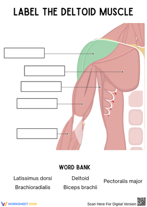
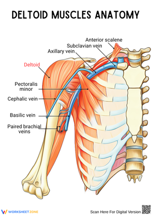
The labeling worksheet presents anterior and posterior shoulder diagrams with blank leader lines pointing to each deltoid head, adjacent structures (trapezius, supraspinatus, teres minor), and key bony landmarks. The function-matching worksheet pairs each head with its primary movement and includes deliberate distractors — scapular elevation, elbow flexion — that push students to reason rather than guess. The origin-and-insertion chart presents each head as a partially completed row; students fill in missing cells for origin, insertion, action, and innervation. The clinical-application worksheet pairs a short reading on intramuscular injection sites with comprehension questions and a diagram where students mark the safe injection zone. The movement-arrows worksheet asks students to draw directional vectors on a standing figure indicating which anatomical plane each head's action occurs in — a task that connects static diagram work to the language of kinesiology. The final comparison worksheet sets the deltoid against the four rotator cuff muscles in a structured table covering origin, insertion, primary action, nerve supply, and common injury patterns.
Student Errors That Surface Fast — and What to Do About Them
The most consistent error: students conflate the middle head with the entire muscle and write "abduction" as the deltoid's only function. Ask them what the anterior head does and they either leave the cell blank or write "lifts the arm" — a description too vague to distinguish flexion from anything else. This happens because introductory instruction often leads with the deltoid's silhouette and tags abduction as its signature action, which students encode as the whole story. The function-matching and movement-arrows worksheets directly address this by requiring students to assign specific movements to each head in isolation.
On the origin-and-insertion chart, students regularly write "scapula" as the origin for all three heads because they know the deltoid connects to bones in the shoulder girdle region. The chart forces exactness — lateral third of the clavicle for the anterior head, acromion process for the middle, spine of the scapula for the posterior — and that precision is exactly what lab practical questions test. A third error surfaces around nerve identification: students frequently substitute the radial nerve for the axillary nerve, particularly after learning that the radial nerve runs through the radial groove of the adjacent humerus. Short-answer prompts requiring the nerve name and spinal levels (C5–C6) catch this substitution before the unit exam.
Building These Worksheets Into Your Anatomy Unit
Sequencing matters more than most teachers expect with deltoid content. Handing out the origin-and-insertion chart on the first day typically produces blank stares, because students have no visual frame yet for where each head sits relative to the others. Start with the labeling worksheet, and pair it with a brief self-palpation moment: have students rest one hand on the opposite shoulder and work through abduction, flexion, and extension to feel which portion of the deltoid engages during each movement. That tactile experience gives them a reference no diagram alone provides, and the chart and matching activities the following day are noticeably more productive for it.
The deltoid muscle worksheets sequence naturally across a five-day shoulder unit. Day one covers labeling and palpation. Day two brings the origin-and-insertion chart as guided practice with teacher modeling at the board. Day three moves to function-matching and movement-arrows as independent work. Day four introduces the clinical-application worksheet — especially valuable in health science CTE courses where injection technique appears later in the year. Day five uses the rotator cuff comparison as a review activity or extension for students who finish early. The clinical worksheet earns a specific caution: introduce it only after students have the three heads secure. Students who attempt to mark the safe injection zone without that foundation often draw marks that would clip the axillary nerve — an error worth naming explicitly during whole-class debrief.
Adjusting Each Worksheet for Different Student Readiness Levels
Adapting these deltoid muscle worksheets for different readiness levels rarely requires creating new materials — it usually means adjusting the prompt or removing a support structure already built into the task. For the labeling worksheet, providing a word bank shifts the task from pure recall to recognition, which reduces frustration for students still anchoring the vocabulary without eliminating the spatial reasoning demanded by leader lines on a diagram. For students working ahead, remove the word bank entirely and add a second layer: after labeling, write one sentence explaining why each head's origin makes mechanical sense given its action. Why does it follow that the anterior head — whose job is shoulder flexion — originates on the clavicle rather than the spine of the scapula?
The comparison table runs at an advanced level by design, but teachers can bring it within reach for mixed-ability classes by pre-filling the rotator cuff rows and asking students to complete only the deltoid columns. This keeps the full comparative structure intact while reducing the total volume of new recall required at once. For students in health science or allied health pathways, the clinical-application worksheet extends further without any new materials: ask them to add a written rationale column to the injection diagram and research two contraindications for deltoid injection sites. The extension requires only a more demanding prompt written at the top of that same worksheet.
Frequently Asked Questions
Do these worksheets suit both high school biology and health science CTE courses?
The labeling, chart, and matching worksheets carry well across both contexts, though the emphasis shifts. Biology courses typically treat the clinical worksheet as enrichment. Health science CTE teachers often make it core content, since injection site anatomy appears later in patient-care and clinical-skills curriculum. The comparison table bridges particularly well to physical therapy and sports medicine pathways where rotator cuff injury is a recurring topic.
What anatomy vocabulary should students know before starting?
Students should be comfortable with anatomical planes of movement, directional terms, and the origin-vs.-insertion distinction before tackling the chart or movement-arrows worksheet. The labeling worksheet works as a reasonable entry point for students still building that vocabulary — particularly when paired with a one-page anatomical terms reference rather than a full textbook glossary.
Are these worksheets appropriate for AP Biology?
AP Biology does not cover musculoskeletal anatomy at this level of specificity, so these deltoid muscle worksheets fit anatomy and physiology electives, health science CTE pathways, and introductory kinesiology courses most directly. AP Biology teachers who want a deltoid-specific case study for motor unit recruitment or nerve innervation concepts can adapt the short-answer and chart components, but the full set maps most cleanly onto dedicated anatomy courses.
How long does each worksheet take during a class period?
The labeling and function-matching worksheets each run 10–15 minutes as independent work, which fits comfortably inside the practice block of a 50-minute period. The origin-and-insertion chart takes closer to 20 minutes for most students working without reference materials. The clinical-application and comparison worksheets run 25–30 minutes and work better as homework with a structured in-class debrief, or during an 80-minute block period.