11th Grade Foot Muscle Printable Worksheets
These 11th grade foot muscle printable worksheets give anatomy teachers direct access to one of the most structurally demanding topics in high school life science. The human foot packs twenty-six bones, thirty-three joints, and well over a hundred individual muscles, tendons, and ligaments into a region students have to understand not just by name but by functional layer — and by junior year, identification alone is no longer enough. This set moves students from recognition to reasoning.
Extrinsic and Intrinsic Muscles: The Core Distinction
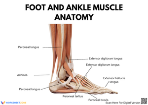
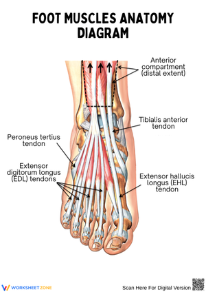
The organizing framework running through this set is the extrinsic-intrinsic classification. Extrinsic muscles — the gastrocnemius, soleus, tibialis anterior, fibularis longus, and their compartment neighbors — originate in the lower leg and act on the foot through long tendons. They drive dorsiflexion, plantarflexion, inversion, and eversion: the large, powerful movements involved in walking, running, and quick direction changes. Students label origin and insertion points, trace tendon paths across the ankle, and match each muscle to its compartment in the lower leg.
Intrinsic muscles present a different kind of problem. They originate and insert entirely within the foot, handling fine motor adjustments — stabilizing the toes during push-off, maintaining arch height under load, and making the continuous micro-corrections that keep a person balanced on uneven ground. Each worksheet covering intrinsic anatomy asks students to work with the four plantar layers separately before synthesizing them, naming muscles, tracing functional relationships, and explaining why layer depth maps onto task specificity.
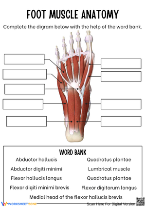
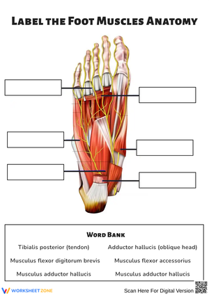
The Four Plantar Layers in Detail
The layered structure of the plantar surface is where students typically hit their first real wall. Standard textbook diagrams that show all four layers simultaneously tend to produce frustrated guessing rather than genuine understanding — there are simply too many overlapping structures in too small a space. These worksheets address that by isolating each layer before asking students to integrate them.
- First layer (superficial): Students work with the abductor hallucis, flexor digitorum brevis, and abductor digiti minimi — the muscles most directly involved in basic toe movement and medial arch support during weight-bearing.
- Second layer: The quadratus plantae and four lumbricals come into focus here. A consistent sticking point is understanding why the quadratus plantae exists at all — specifically, how it corrects the oblique pull of the flexor digitorum longus tendon so that the toes flex straight rather than diagonally. Each worksheet in this layer asks students to diagram that correction explicitly.
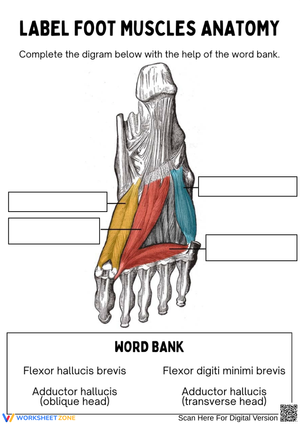
- Third layer: The flexor hallucis brevis, adductor hallucis, and flexor digiti minimi brevis — muscles that give the big toe and little toe their independent control during balance adjustments and late-stance propulsion.
- Fourth layer (deepest): The dorsal and plantar interossei, responsible for toe abduction and adduction respectively. Students frequently reverse these, which is why this worksheet includes a built-in error-correction prompt that asks them to write out the logic before labeling.
The 11th grade foot muscle printable worksheets in the plantar series use a warm-to-cool color-coding system — warm tones for the first and second layers, cool tones for the third and fourth — which maps depth visually and reduces the cognitive load of tracking which muscle belongs where. Students who use the color system consistently perform measurably better on layer-identification assessments than those who label in a single pen color.
Connecting Anatomy to Function and Clinical Context
Anatomical facts are embedded in functional questions throughout this set rather than isolated in pure identification tasks. One worksheet presents a runner with pain along the medial longitudinal arch and asks students to identify which intrinsic muscles are most likely involved and what their relationship is to the plantar aponeurosis. Another asks students to explain what mechanical problem the quadratus plantae solves and what would happen to toe flexion if it were absent. These are not hypothetical scenarios — they describe the kinds of questions that appear in sports medicine, physical therapy, and college-level anatomy courses.
Many Grade 11 students have experienced plantar fasciitis, shin splints, or ankle sprains personally or through a teammate, and that lived experience gives the vocabulary real traction. When students can connect the abductor hallucis to a condition they have actually felt, the Latin root stops being an obstacle and starts being a useful label.
The Errors That Appear Most Often in Student Work
The most persistent mistake in Grade 11 foot anatomy is conflating tendon path with muscle origin. Students correctly understand that the flexor digitorum longus is an extrinsic muscle — it lives in the posterior compartment of the lower leg — but then label its tendons, which run through the sole of the foot, as intrinsic muscle structures. The extrinsic worksheets address this with tracing exercises that require students to mark both the muscle belly in the leg and the tendon insertion in the foot on the same diagram, forcing them to hold both ends of the structure in view simultaneously.
Layer confusion in the plantar surface is the other consistent problem. Students who memorize muscle names in isolation frequently place them in the wrong layer because they have no spatial image of the depth involved. The lumbrical error is especially predictable: students almost always assign the lumbricals to the first layer because the lumbricals feel like basic toe-flexors conceptually. In reality, the lumbrical tendons travel beneath the deep transverse metatarsal ligament and pass dorsal to it — a detail that changes their mechanical function entirely. The second-layer worksheet includes a cross-section diagram precisely to anchor that three-dimensional relationship before students attempt to label the plantar surface from above.
Building These Worksheets Into Your Weekly Anatomy Block
Most anatomy teachers using this set find the most efficient entry point is the extrinsic muscles worksheet as a pre-lab primer — either the class period before a lower-limb model lab or as a homework assignment students complete before dissection day. When students arrive already knowing origin, compartment, and primary action for the major extrinsic muscles, the hands-on work can focus on observation and confirmation rather than first exposure, which makes the limited lab time far more productive.
The plantar layer worksheets work well as a station rotation. Set up four stations with a plastic foot model, a cadaver atlas open to each layer, the corresponding worksheet, and a brief written task. Students move through in small groups, using the worksheet to check what they observe against labeled anatomy. The color-coding system transfers directly to the model — students can mark depth with colored dot stickers the same way the worksheets code it, and that physical act of placing stickers reinforces the layering better than re-reading a diagram does.
The clinical case worksheets function well as Friday application tasks after a week of identification work. They ask students to pull together three or four concepts at once — arch structure, muscle function, tensile load, and injury mechanism — which makes them stronger summative checks than labeling exercises alone. Distribute them at the end of the unit sequence, not midway through.
Standard Alignment
This set aligns with NGSS HS-LS1-2 (From Molecules to Organisms: Structures and Processes), which asks students to develop and use models to illustrate the hierarchical organization of interacting body systems. In a foot anatomy unit, that standard shows up concretely when students must explain not just what a muscle is named but how it interacts with tendons, bones, and sensory nerves to produce coordinated movement. A task asking students to trace the feedback loop between proprioceptive nerve endings in the intrinsic muscles and the motor response that adjusts arch height is working directly against that standard — not just covering anatomy as a naming exercise.
Differentiating Across Learner Levels in a Mixed Anatomy Class
Students who are still building their anatomical vocabulary benefit from starting with the extrinsic muscle worksheets, since those muscles are larger, fewer in number, and connected to movement patterns students already understand intuitively — everyone knows what the calf does. Starting with the gastrocnemius-soleus group and expanding outward to the anterior and lateral compartments gives those students a functional anchor before they encounter the intrinsic muscles, where the names are unfamiliar and the movements are subtle.
For students moving toward pre-medical electives or Advanced Placement Biology, the clinical case worksheets support genuine extension. Ask them to research the conservative treatment protocol for plantar fasciitis and then annotate their completed worksheet to show which specific muscles are targeted during each phase of rehabilitation — from initial rest and arch taping through progressive loading and eccentric strengthening. That kind of task turns a labeling exercise into biomechanical reasoning that transfers directly to college-level anatomy coursework. The 11th grade foot muscle printable worksheets in the clinical section include enough anatomical detail to support that depth without requiring additional references.
Frequently Asked Questions
Which muscles should I prioritize if class time is limited?
Start with the extrinsic group: tibialis anterior, tibialis posterior, fibularis longus and brevis, gastrocnemius, and soleus. These are the muscles students encounter most often in movement science, sports medicine contexts, and college anatomy prerequisites. From the intrinsic group, prioritize the first plantar layer and the interossei — those two groups account for the majority of assessment questions at this level, and they anchor the functional concepts that make the middle layers easier to understand later.
Do these worksheets require a physical model to use effectively?
No. The cross-section diagrams and layer-by-layer format let students work productively from the worksheets alone. That said, pairing even a single class session with a plastic foot model or a cadaver atlas image meaningfully improves retention of the layered relationships — the worksheets include spatial reference points that transfer directly to a model when one is available, so the two resources reinforce rather than duplicate each other.
Can these be used with students who are still behind in anatomical terminology?
The 11th grade foot muscle printable worksheets in the extrinsic section include a reference glossary of directional terms and movement vocabulary printed on the same worksheet, so students do not need to flip between resources during the task. For students who are significantly behind in terminology, the glossary provides enough support to complete the labeling work independently while still meeting the core learning objectives for the unit.
Clear All