11th Grade Calf Muscle Worksheets (PDF)
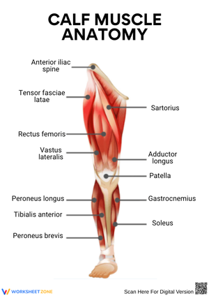
These calf muscle worksheets pdf for 11th grade give anatomy teachers a focused set of resources for the posterior lower leg — a unit that demands more spatial reasoning than students expect, because the gastrocnemius and soleus overlap in both location and function, and keeping them distinct requires repeated, deliberate exposure.
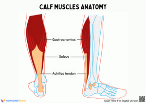
The Specific Anatomy and Physiology Covered
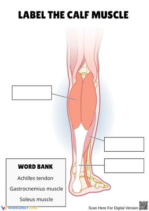
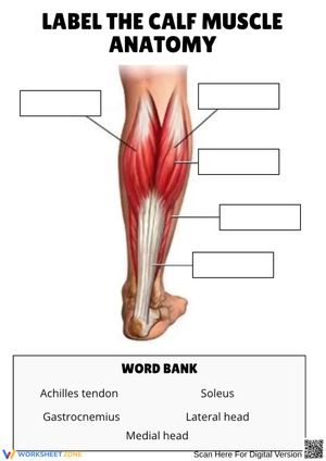
The core focus is the triceps surae — the gastrocnemius and soleus together — and how they converge into the calcaneal tendon. Each worksheet addresses the anatomical content at a different level of depth, moving from basic identification to functional analysis to clinical application. Across the set, students work through:
- Labeling the medial and lateral heads of the gastrocnemius and their origins on the posterior femoral condyles
- Identifying the soleus origin along the posterior tibia and fibula, distinct from and deeper than the gastrocnemius
- Tracing the Achilles tendon from its muscular origins to the calcaneus and explaining why it is considered the strongest tendon in the body
- Classifying the ankle-heel system as a second-class lever, with the ball of the foot as the fulcrum, body weight as the load, and calf contraction as the effort force
- Comparing the slow-twitch fiber dominance of the soleus against the mixed fiber composition of the gastrocnemius, then connecting fiber type to specific athletic demands
- Applying posterior leg anatomy to clinical scenarios: Achilles tendinopathy, gastrocnemius mid-belly strain, and the skeletal muscle pump's role in venous return
That last item — the venous pump — tends to surprise students the most. The idea that the calf muscles act as a secondary circulatory engine during walking, compressing deep veins and pushing blood upward against gravity, connects musculoskeletal anatomy to cardiovascular physiology in a way most students have not encountered before.
Errors That Come Up Repeatedly in Student Work
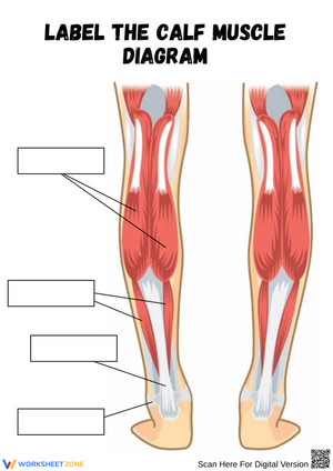
The most consistent mistake is misplacing the soleus origin. Students who correctly label the gastrocnemius heads on the femoral condyles will often draw the soleus originating from the same region on a blank diagram, erasing the anatomical distinction that determines whether a muscle can act on the knee at all. The calf muscle worksheets pdf for 11th grade address this directly through labeling tasks that require students to name not just the muscle but the specific bone of attachment — an answer that changes the entire functional analysis that follows.
A second predictable error involves the biarticular principle. When asked which muscle is more active during a seated calf raise — knee bent at 90 degrees — most students answer "gastrocnemius" on the assumption that the larger, more visible muscle dominates in every position. Working through the functional questions on these worksheets forces students to reason from origin location to joint mechanics: a gastrocnemius shortened at the knee loses mechanical advantage at the ankle, which means the soleus carries the load in that position. Students who arrive at that conclusion through their own reasoning retain it; students told the rule without the reasoning forget it by the next class.
Plantar flexion and dorsiflexion also get reversed on movement diagrams more often than expected, particularly when the diagram shows the foot from an unfamiliar lateral angle. A simple anchor helps: dorsiflexion moves the toes toward the shin, parallel to how the word "dorsal" describes the top surface of the hand or foot. Students who connect the term to a visual reference stop reversing the two consistently.
Sequencing These Worksheets Through Your Posterior Leg Unit
These calf muscle worksheets pdf for 11th grade are most effective when the blank labeling task comes before formal lecture rather than after. Students who encounter an unlabeled posterior leg diagram without pre-teaching generate specific questions about what they cannot identify — which reduces the cognitive load required to encode the answer when instruction follows. That inversion of the standard lecture-then-practice sequence takes confidence to execute the first time, but the quality of student questions during the debrief consistently justifies it.
The calf raise palpation activity pairs directly with the fiber-type comparison worksheet. Have students stand at their desks and perform a slow heel raise with knees straight, hold for three seconds, then repeat with knees bent to about 30 degrees. The gastrocnemius softens noticeably in the bent-knee position as the soleus takes over — students can feel the shift through their own hand placed on the back of their calf. Completing the Type I versus Type II fiber comparison worksheet immediately after anchors the histological distinction to something they just experienced physically rather than read about abstractly.
For the clinical scenario worksheets, small-group work outperforms independent silent completion. Assign each group a different injury — Achilles rupture, mid-belly gastrocnemius strain, soleus overuse from distance running — and ask them to explain the anatomical mechanism using their labeled diagrams. Give each group one sentence to present at the end of class, then correct any misidentified structures in real time. Groups that must reason aloud expose gaps that written answers would mask.
Standard Alignment
These resources align with NGSS HS-LS1-2, which addresses how the hierarchical organization of interacting systems provides specific functions within multicellular organisms. The progression built into this set — from muscle fiber histology through whole-muscle mechanics and into organ-system interaction via the venous pump — maps directly onto that standard's requirement for multi-scale analysis. In most 11th-grade anatomy sequences, HS-LS1-2 appears mid-year during the musculoskeletal unit, placing these worksheets naturally in the second or third quarter, after students have covered basic tissue types but before the nervous system unit begins.
Making the Set Accessible Across Ability Levels
The calf muscle worksheets pdf for 11th grade include labeled reference diagrams alongside the blank labeling tasks, which gives teachers a straightforward way to tier the work. Students who need more support use the reference diagram as an open resource during the labeling worksheet; students working at grade level complete the blank version first, then check accuracy against the reference afterward. For students who freeze when encountering an unfamiliar posterior view of the leg — a common response when the diagram angle differs from the textbook illustration — adding a brief orientation note ("medial side on the right, lateral on the left") removes the visual ambiguity without reducing the anatomical challenge.
Advanced students benefit from moving past identification into quantitative application. Have them estimate the mechanical advantage of the calf's lever system using measurements from their own foot — distance from heel to ball of foot as the effort arm, distance from heel to ankle joint as the load arm — and compare the result to a lever elsewhere in the body. Students who find the labeling procedurally easy generally engage with calculation-based extensions more readily than with additional anatomical detail at a finer level, because the cognitive challenge shifts from recall to analysis.
Frequently Asked Questions
What distinguishes the function of the gastrocnemius from the soleus?
Both muscles produce plantar flexion of the ankle, but the gastrocnemius also assists in knee flexion because its origin sits above the knee joint on the femoral condyles. The soleus only crosses the ankle. Their fiber compositions also differ: the gastrocnemius carries a higher proportion of fast-twitch fibers for explosive movement, while the soleus is predominantly slow-twitch — suited to the sustained, fatigue-resistant work of maintaining standing posture throughout the day.
How does the calf muscle pump assist in venous circulation?
The deep veins of the lower leg run through the tissue surrounding the posterior leg muscles. When the gastrocnemius and soleus contract during walking or any weight-bearing activity, they compress those veins and push blood upward against gravity. One-way valves inside the veins prevent backflow when the muscles relax, producing a rhythmic pumping action. Prolonged immobility — sitting through a long flight, for example — reduces this pumping action, which is one reason extended inactivity raises the risk of deep vein thrombosis.
Which nerve supplies the posterior leg muscles?
The gastrocnemius, soleus, and the rest of the posterior compartment muscles are innervated by the tibial nerve, a major branch of the sciatic nerve that travels down the back of the thigh and leg. The tibial nerve carries both motor fibers controlling plantar flexion and sensory fibers serving the sole of the foot, so a tibial nerve injury affects movement and sensation in the same region simultaneously.
Why does bending the knee reduce the gastrocnemius's contribution to plantar flexion?
Because the gastrocnemius originates above the knee, flexing the knee shortens the muscle before it acts on the ankle joint. A shortened muscle on the length-tension curve generates less force than one at optimal resting length, so the gastrocnemius contributes far less to plantar flexion when the knee is flexed. Seated calf raise exercises take advantage of this principle — the bent-knee position reduces gastrocnemius involvement and forces the soleus to do the primary work, which is why physical therapists use that position to isolate the soleus during rehabilitation.
Clear All