11th grade deltoid muscle worksheets printable give anatomy teachers a concrete way to push students past passive reading and into genuine structural analysis of how the shoulder moves. Each worksheet in the set targets the deltoid's three fiber groups, their individual origin points, and the way they converge at a single insertion to drive a wide range of arm movements. Teachers get labeled diagrams, origin-insertion-action tables, and short clinical application prompts — the kind of material that builds the working vocabulary students need before a dissection or model lab.
What Each Worksheet Covers
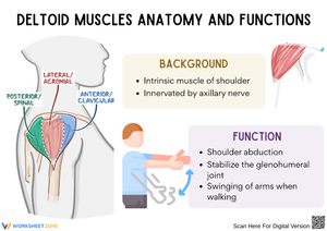
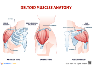
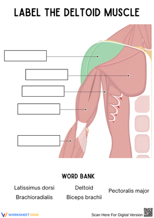
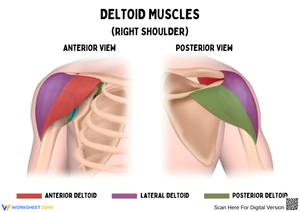
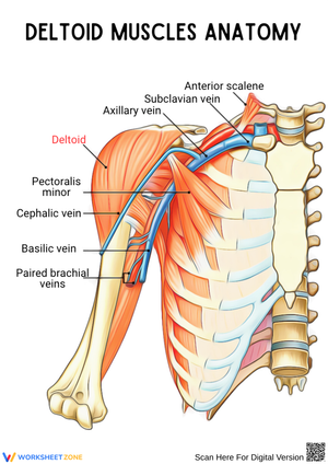
The set addresses the deltoid as a divided structure, not a single muscle mass. Students identify the anterior (clavicular) fibers, originating at the lateral third of the clavicle; the middle (acromial) fibers, originating at the acromion of the scapula; and the posterior (spinal) fibers, originating along the spine of the scapula. Each fiber group inserts at the deltoid tuberosity on the lateral humerus. Students practice tracing those convergent force lines from three separate starting points to one shared endpoint — an exercise that makes the muscle's mechanical versatility visible on paper before they encounter it in a model or specimen.
Beyond labeling, students work with OIA tables that require them to write out the specific actions each head performs. The anterior fibers drive flexion and medial rotation; the middle fibers execute abduction above 15 degrees of arm movement; the posterior fibers produce extension and lateral rotation. Documenting all six actions across three heads forces students to think in columns rather than treating the deltoid as one entity with one job. Each worksheet also includes a synergist-and-antagonist prompt, asking students to identify the supraspinatus as the initiator of abduction and the latissimus dorsi as an opposing force in certain rotational movements.
Errors That Show Up Repeatedly in Student Work
The most consistent mistake is swapping the anterior and middle heads when labeling diagrams. Students who correctly memorize "clavicle = anterior" will still circle the wrong region because they anchor the clavicle to the midline of the chest rather than its lateral third. The error is spatial, not vocabulary-based. Having students trace the lateral clavicle with a finger on their own collarbone before labeling the diagram corrects this faster than re-reading the definition.
A second persistent error involves the insertion description. Students regularly write "deltoid tuberosity on the humerus" and omit the qualifier "lateral aspect." That omission matters for understanding force vectors — if the tuberosity were medial, abduction would not be mechanically possible. Students who connect insertion location to function retain the detail through exam day; those who only memorize the name lose it by the following week. Marking insertion latitude directly on the bone diagram, rather than just writing the name beside an arrow, closes that gap reliably.
Fitting These Worksheets Into Your Anatomy Unit
11th grade deltoid muscle worksheets printable work best as structured preparation before hands-on work, not as post-lab review. When students complete an OIA table and a fiber-labeling diagram before they handle an anatomical model or palpate a partner's shoulder during a movement lab, the hands-on session becomes a confirmation exercise rather than an orientation session. That shift in cognitive load frees up lab time for higher-order questions — about force direction, clinical implications, and muscle failure modes — instead of basic identification.
The resistance-band extension is worth building into at least one class period. Have students perform lateral raises with a light band while annotating their labeled diagrams: mark which head is the prime mover, which is stabilizing, and which is largely inactive. A front raise shifts activity to the anterior fibers, and students annotate accordingly. Comparing two movement patterns on the same worksheet makes muscle fiber specificity tangible in a way that colored diagrams alone cannot replicate. A 10-minute version of this works during the block period after students have finished the written portion of each worksheet.
As a warm-up format, one worksheet per class at the start of the shoulder unit provides spaced retrieval practice before the unit exam. The 8 minutes between the bell and the start of direct instruction is enough time to complete a short OIA prompt without cutting into lesson delivery.
Standard Alignment
These worksheets align with NGSS HS-LS1-2, which asks students to develop and use models to illustrate the hierarchical organization of interacting systems. The deltoid is an unusually clean fit for that standard: students model how three skeletal attachment points on the same muscle determine three distinct mechanical outcomes. The standard calls for function-level analysis, not just identification, and the OIA table format pushes students directly toward that kind of thinking. At the AP Biology level, this material also supports the enduring understanding that structure and function are inseparable across biological scales.
Adjusting the Set for Different Levels of Student Preparation
11th grade deltoid muscle worksheets printable in this set include both partially labeled diagrams with word banks and fully blank versions. Students new to anatomical terminology do better starting with the word bank and a partially completed OIA table, then moving to the blank versions once the vocabulary is stable. Students with prior anatomy coursework — or a semester of sports medicine behind them — can skip the word bank version entirely and use the completed worksheet as a self-check afterward.
For students who struggle with the three-dimensional geometry of the shoulder, a simple overhead-view sketch of the clavicle, acromion, and scapular spine placed alongside the standard anterior diagram significantly reduces confusion about origin locations. That sketch takes two minutes to draw on the board and is worth doing for any class where more than three students are consistently mislabeling the fiber origins. It does not replace the worksheet — it makes the worksheet more productive for students who need spatial orientation before they can label accurately.
Frequently Asked Questions
Do these worksheets address the axillary nerve?
Yes. One worksheet includes a prompt on the axillary nerve (C5–C6) as the deltoid's primary innervation, with a short clinical note about the consequences of a fracture at the surgical neck of the humerus or a shoulder dislocation. Students mark the nerve's approximate path on the diagram and answer an inference question about why deltoid atrophy follows specific shoulder injuries. That clinical prompt makes the anatomy feel consequential rather than purely taxonomic.
Are these suitable for AP-level courses, or only standard 11th grade biology?
Both. The core labeling and OIA exercises fit standard anatomy or biology courses. The synergist-antagonist analysis and the clinical injection-site application extend naturally into AP Biology and AP Human Anatomy, where structure-to-function reasoning drives most of the exam questions. In mixed classes, teachers assign the core diagrams to all students and the clinical extension prompts to those working at the advanced level.
How do these worksheets support exam preparation?
11th grade deltoid muscle worksheets printable build active recall through repeated labeling and table completion — the same retrieval format anatomy exams use. Students who have filled in OIA tables multiple times recognize the question format immediately and retrieve the answer rather than reconstructing it under pressure. The clinical application prompts also prepare students for application-level questions, which appear more frequently on standardized assessments than simple identification tasks do.