These spinal cord printable worksheets give biology teachers a focused set of activities that move students from basic anatomical labeling into functional reasoning — the structure-function connection at the core of any nervous system unit. Each worksheet in the set targets a discrete skill: cross-section identification, meningeal layer sequencing, reflex arc pathway tracing, or regional division mapping. Together they cover the anatomy content that recurs on both unit tests and state science assessments in grades 6–12.
The Specific Skills Targeted
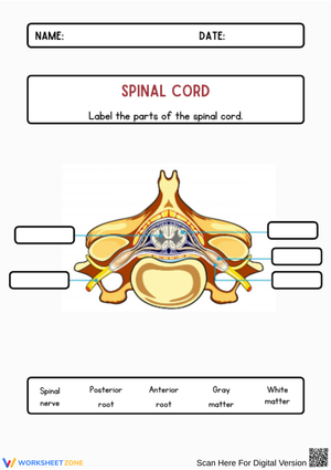
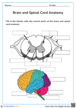
Four content strands run through the collection. Cross-section labeling asks students to mark the butterfly-shaped gray matter interior and the surrounding white matter tracts, then identify the dorsal root, ventral root, central canal, and dorsal root ganglion on the same diagram. The gray matter / white matter distinction is foundational — gray for neuronal cell bodies and interneurons, white for myelinated ascending and descending axon tracts — and students who blur it early misread every structure-function question that follows.
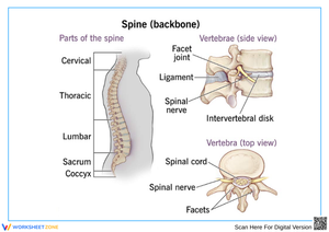
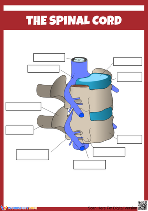
Reflex arc worksheets trace the five-component pathway: sensory receptor → afferent neuron → spinal cord interneuron → efferent neuron → effector muscle. Students annotate each node with signal direction and neuron type, which forces them to treat the pathway as a sequence rather than a static vocabulary list. Protective structure worksheets cover the three meningeal membranes (dura mater, arachnoid mater, pia mater), cerebrospinal fluid, and the vertebral column; students sequence these layers from skin inward, building spatial understanding that recall-based questions alone don't develop. Regional division worksheets pair a longitudinal diagram with activities that connect each spinal segment — cervical, thoracic, lumbar, sacral, coccygeal — to the body areas it controls, followed by short injury-level case prompts that anchor abstract segment names in clinical reasoning.
Frequent Student Errors Worth Watching For
The gray matter / white matter reversal persists well past the initial lecture. Students who can repeat the definitions correctly will still locate the regions incorrectly on a diagram — they hear "gray matter is inside" but when they encounter a cross-section where the butterfly shape is drawn with a light fill on white paper, the visual doesn't reinforce the name. Color-coding both regions with pencil before writing any labels reduces this error more reliably than re-explanation does.
Reflex arc errors cluster at the afferent/efferent junction. Students who define "afferent" correctly in a word bank will still draw the signal arrow pointing away from the spinal cord on the afferent neuron, because they're working from verbal recall rather than spatial reasoning about the pathway. Worksheets that require directional arrows alongside neuron labels — not just vocabulary fill-ins — surface this error before it appears on a test.
A subtler pattern involves the spinal roots. Many students memorize "dorsal = sensory, ventral = motor" but cannot apply that to an unlabeled diagram when the cross-section is rotated from the orientation they first encountered. Varying diagram orientation across the set addresses this more efficiently than any amount of re-teaching the rule.
How to Sequence These Worksheets Across a Unit
Start with the longitudinal diagram worksheet before any cross-section work. Students who encounter the cord as a floating anatomical structure — disconnected from the vertebral column and the body — consistently misapply the regional division content later. That orientation step is not optional. Cross-section labeling follows the next day, done in pairs with a whole-class comparison after. The pair-check is particularly productive here because students argue about dorsal versus ventral root placement in ways that a teacher explanation doesn't provoke.
The reflex arc worksheet lands best after a live demonstration rather than before it. Test a student's patellar reflex in front of the class — it takes ninety seconds — then distribute the flowchart worksheet immediately. Students who have just watched the reflex happen are far more willing to commit to directional arrows on the pathway diagram. One retrieval move worth building in: the morning after students complete the spinal cord printable worksheets for the reflex arc, open class with a three-minute blank-paper redraw — no notes, no diagram in front of them. The errors that appear reveal exactly which node in the sequence hasn't solidified, and the exercise takes less time to administer than any formal quiz.
Standard Alignment
These materials align most directly with NGSS performance expectation HS-LS1-2, which asks students to develop and use models to illustrate how cellular structures enable organ-level functions — a framing that maps cleanly onto the gray matter / white matter architecture and the reflex arc pathway. For middle school courses under MS-LS1-3, the structure-function connection is the same standard applied at a less mechanistic level of detail. In both cases, the labeling and pathway worksheets serve as the model-development activity the standard describes; injury-level case prompts push students toward the evidence-based explanation component. Teachers in states with separate anatomy and physiology standards will typically find the set aligns to the nervous system strand, usually in the same instructional unit as brain and peripheral nervous system content.
Adjusting the Worksheets for Different Student Levels
For students who need more support, the cross-section labeling worksheet works well with a partially completed word bank — provide four of the six structure labels and let students determine placement for the remaining two. This narrows the cognitive load on identification without eliminating the reasoning step. Protective layer matching activities can be modified by pre-drawing the connecting lines and asking students to annotate the function of each layer, shifting the demand from retrieval to comprehension.
Advanced students benefit from extension prompts that move beyond identification. After completing the regional division worksheet, ask them to write a clinical prediction: if the cord were compressed at the T6 vertebral level, which motor and sensory functions would be disrupted, and why? That prompt requires students to integrate regional anatomy, gray/white matter knowledge, and ascending/descending tract content simultaneously — a strong formative task for students who race through labeling activities. These spinal cord printable worksheets are written at a level of anatomical detail that supports this kind of extension without requiring a separate document; the core diagrams give advanced students enough information to push deeper on the same worksheet they're already using.
Frequently Asked Questions
What is the difference between gray matter and white matter in the spinal cord?
Gray matter forms the butterfly-shaped inner region and contains neuronal cell bodies and the interneurons that process incoming signals. White matter surrounds it and consists of myelinated axon tracts — organized bundles carrying sensory information toward the brain (ascending) and motor commands away from the brain (descending). Students who only read the definitions tend to reverse the locations under pressure; those who color-code the regions before labeling retain the distinction significantly better.
How does a spinal reflex produce movement before the brain responds?
The signal from the receptor travels along an afferent neuron to an interneuron housed within the spinal cord. That interneuron activates the efferent motor neuron directly, triggering the effector muscle without waiting for the signal to ascend to the cortex and return. The brain receives notification afterward — which is why the reflex is consciously felt — but the protective movement completes first. The spinal cord printable worksheets for the reflex arc pathway include directional arrows at each step, which makes the bypass mechanism concrete rather than abstract.
What are the five spinal cord regions and which body areas do they control?
The cervical region (C1–C8) controls the neck, shoulders, arms, and hands. The thoracic region (T1–T12) governs chest muscles and the upper abdomen. The lumbar region (L1–L5) controls the lower back, hips, and legs. The sacral region (S1–S5) manages the pelvis, bladder, bowel function, and lower limb reflexes. The small coccygeal segment provides limited cutaneous innervation at the tailbone. Regional diagram worksheets that ask students to draw connecting lines between each segment and its corresponding body areas build this knowledge more durably than flashcard review.
At what grade level are these worksheets appropriate?
Cross-section labeling and protective layer worksheets work well in grades 6–8 life science when the instructional focus stays on structural vocabulary without requiring mechanistic explanation. Reflex arc and regional division content suits grades 9–12 biology, anatomy and physiology, or health science courses, where students are expected to explain mechanism rather than simply identify structure. Teachers typically select individual worksheets based on course level rather than working through the full set in sequence.