These knee worksheets pdf resources give secondary anatomy, health science, and sports medicine teachers a ready-to-print entry point into one of the most structurally demanding joints students will study. The set covers bony landmarks, stabilizing ligaments, cartilage structures, and tendons — the full roster of content that appears on every major anatomy assessment at this level. Each worksheet targets a specific skill, so teachers can assign them individually or sequence them across a unit without overlap.
Structures Targeted Across the Set
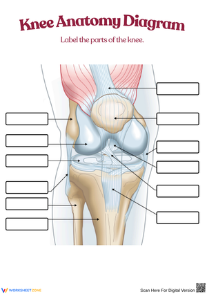
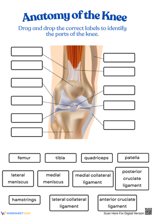
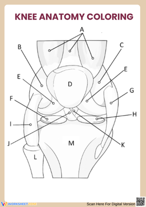
The knee sits at the intersection of the femur, tibia, and patella, with the fibula contributing laterally as an attachment point for the LCL. Students need to hold roughly twelve distinct structures in working memory before they can reason about injury, function, or clinical context. These worksheets break that load into manageable pieces: one worksheet isolates bony landmarks, another focuses on the four major ligaments, and a third introduces cartilage and bursae together — limiting each task to a focused subset rather than asking students to process everything at once.
The four ligaments are consistently the heaviest lift. The ACL and PCL are named for their tibial attachment sites — anterior and posterior — not their femoral origins, and that distinction trips up roughly half the class on first contact. Each ligament worksheet asks students to identify attachment points and state the movement each ligament resists, which builds functional logic rather than isolated term recall. The menisci are handled through a superior view of the tibial plateau, a perspective that makes the C-shape and relative positioning far clearer than a standard lateral illustration.
Frequent Mistakes That Show Up When Students Label the Knee
The ligament-tendon distinction produces the most consistent errors at this level. Students who can define both terms on a vocabulary quiz will still write "patellar ligament" on a labeled diagram — they have encountered that phrasing in athletic reporting and it overrides the anatomical definition the moment they are working quickly. The patellar tendon connects the patella to the tibial tuberosity and transmits quadriceps force; it is definitionally a tendon, not a ligament. Running a brief matching activity before the labeling worksheet catches this before it becomes a test-day problem.
A second persistent error involves cruciate ligament direction. Students learn that the ACL prevents anterior tibial translation, but when asked to draw the crossing pattern on a frontal-view diagram, many reverse the X — placing the ACL posterior and the PCL anterior. Asking students to trace each cruciate's fiber path in a different color on the worksheet diagram forces them to follow actual anatomy rather than guess at a label, and the reversal rate drops noticeably after that step.
Building These Worksheets Into Your Lesson Sequence
In a standard anatomy and physiology course, the labeling worksheet works best on the second day of the unit — after students have had one pass through the structures via video or model demonstration, but before the lesson moves to function and injury content. Students who attempt the diagram with no prior exposure tend to fill in terms they have heard from athletic trainers rather than what the illustration actually shows. Using the worksheet as a formative check at that specific point in the sequence keeps pacing tight and surfaces vocabulary gaps early.
Sports medicine teachers find the injury identification activities most effective the week after students read about a current ACL or meniscus injury in the news. The clinical scenario format — students read a mechanism of injury and name the likely damaged structure — lands differently when students have a real athlete in mind. The Friday before a unit test is a natural slot: the activity runs about fifteen minutes, generates enough peer discussion to surface lingering gaps, and requires no additional teacher prep beyond printing.
Adjusting the Set for a Range of Learners
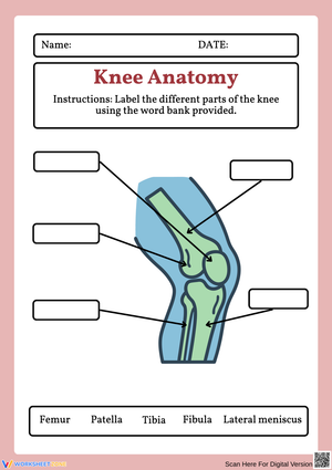
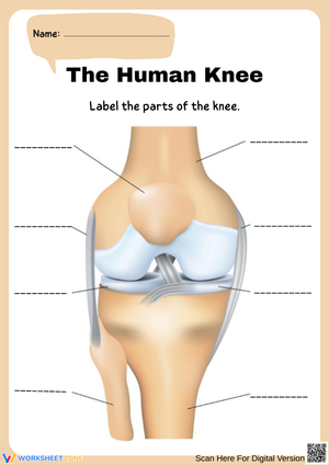
Each knee worksheets pdf download includes a word-bank version and an open-ended version of the same diagram. For students who freeze at a blank illustration, the word bank reduces the retrieval demand enough to let them focus on spatial placement — a separate skill from vocabulary recall, and worth practicing independently. For students who already carry anatomical vocabulary from a prior biology course, the open-ended version pushes them to produce labels without prompting, which often reveals that their mental map of the structures is less precise than they assumed.
Teachers working with mixed-ability classes can run both versions simultaneously. Students who finish the open-ended version early annotate the diagram with functional notes — "resists valgus stress" beside the MCL, "transmits quad force" beside the patellar tendon — extending the task without requiring a separate handout. For English language learners, writing both the English label and a home-language equivalent on the diagram supports retention without undermining the anatomical learning objective.
Standard Alignment
These worksheets align with NGSS HS-LS1-2, which asks students to develop and use a model to illustrate the hierarchical organization of interacting systems in multicellular organisms. At the high school level, the knee is a strong vehicle for this standard: students must reason about how bone, cartilage, connective tissue, and muscle operate as a coordinated unit rather than as isolated parts. State health science and anatomy frameworks within most CTE pathways also address foundational anatomical knowledge as a prerequisite for clinical and therapeutic career credentials. Teachers using the knee worksheets pdf set in a health science elective can document standards coverage in both the science and career-readiness strands without assigning separate activities for each.
Frequently Asked Questions
What structures should students be able to identify for a high school knee anatomy assessment?
The core twelve are the femur, tibia, patella, fibula, ACL, PCL, MCL, LCL, medial meniscus, lateral meniscus, patellar tendon, and quadriceps tendon. Bursae are worth introducing conceptually, but naming individual bursae is typically reserved for post-secondary anatomy courses. Most high school assessments draw from this core list.
Can these worksheets work in a PE or sports medicine class, not just a traditional anatomy course?
Yes — and the injury identification format was built with that context in mind. PE and sports medicine teachers pair the labeling activity with discussions of high school athletic injuries: ACL tears in soccer and basketball, patellar tendinitis in distance runners, meniscus damage in wrestlers and football players. When students can name the structure and connect it to a scenario from their own sport, the anatomical terms stick in a way that diagramming alone does not achieve.
How do these worksheets address the ligament-tendon confusion that shows up in student work?
The matching activity asks students to categorize each structure as ligament or tendon before placing it on the diagram — a deliberate sequencing choice that separates the classification task from the spatial placement task. Teachers who skip the matching step and go straight to labeling consistently see more mislabeling, particularly with the patellar tendon and the collateral ligaments.
Where can I download a printable copy for immediate classroom use?
WorksheetZone offers this complete set as an instant-download collection; each knee worksheets pdf file prints cleanly on standard 8.5 × 11 paper without layout shifts across different printers or operating systems. Teachers can also share the files through Google Classroom, Canvas, or any LMS that supports PDF uploads for digital or hybrid assignments.