




















Foot Worksheets: Printable Human Foot Anatomy Activities for K-12 Science Classes
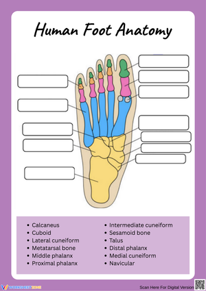
These foot worksheets give science teachers a structured path into one of anatomy's most underestimated topics — the human foot holds 26 bones, 33 joints, and more than 100 muscles, tendons, and ligaments, yet students consistently assume it is simpler than it is until they start labeling it. Each worksheet in the set targets a specific skill: identifying the three bone groups, color-coding the arch system, matching tarsal names to anatomical positions, or connecting structure to function through sentence-completion responses.
Anatomy Content the Worksheets Address
The set is organized around the foot's three anatomical regions: hindfoot, midfoot, and forefoot. Worksheets in each group address the following bone sets:
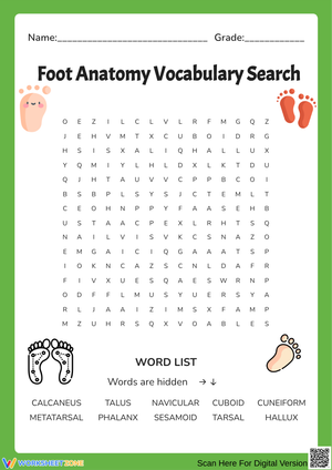
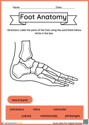
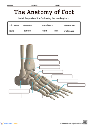
- Tarsals (7 bones): The calcaneus and talus are introduced first at every level. The navicular, cuboid, and three cuneiforms — medial, intermediate, and lateral — appear in activities calibrated for grades 6 and above.
- Metatarsals (5 bones): Each bone is numbered from the hallux (big toe) side inward, with attention to how each bone's position connects to the foot's arch structure and weight-bearing mechanics.
- Phalanges (14 bones): Activities require students to count and label each toe individually — three phalanges for toes two through five (proximal, middle, and distal), two for the hallux — and to explain in writing why the big toe lacks a middle phalange.
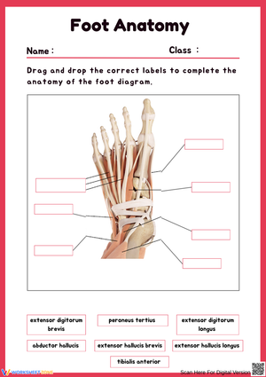
Additional worksheets cover the three arch types: medial longitudinal, lateral longitudinal, and transverse. Each arch activity pairs the labeling diagram with a sentence-completion item asking students to name the arch's specific functional role — so the task goes beyond placing a label to explaining what that structure actually does. Soft tissue worksheets covering the Achilles tendon, plantar fascia, and major flexor and extensor tendon groups are included for high school anatomy and physiology courses and are clearly marked at that level.
Fitting These Worksheets Into Your Lesson Plans
Most teachers run foot anatomy as a four-day embedded sequence inside a skeletal system unit. Day one works best as a vocabulary introduction: a matching activity that pairs bone names with their locations and brief functional notes. Students who attempt a blank labeling diagram without first working through the vocabulary treat the task as guessing rather than anatomy.
Day two's coloring worksheet — shading the tarsal, metatarsal, and phalange groups in three distinct colors — converts the bone-count facts (7, 5, 14) from abstract numbers into a spatial map. The color-separation step does real cognitive work: students who can visually distinguish the three zones before attempting a blank diagram make fewer placement errors and recover faster when they do make them. This is the principle of gradual release applied at the unit scale — coloring first, labeling second.
On day three, foot worksheets work well as a partner activity. Students label independently for ten minutes, then trade papers and check against their coloring sheet from the day before. That self-correction step surfaces errors before they calcify, and it adds nothing to the grading queue. One addition worth building into days three or four, especially in grades four through seven: have students trace their own foot on blank paper, then hold it next to a printed bone diagram at roughly the same scale. The moment a student places a finger on the diagram's calcaneus and recognizes it as the heel of their own traced outline is the kind of concrete anchoring that memorization alone does not produce. The activity takes about eight minutes and consistently reduces the spatial confusion that shows up on labeling quizzes.
Mistakes Students Make That These Worksheets Help You Catch
Two errors appear across grade levels with enough regularity to be worth anticipating before the unit begins. The first is the talus/calcaneus reversal. Students memorize "calcaneus equals heel bone" from notes, but when they encounter the foot in lateral view, they label the talus as the calcaneus because the talus is the more prominent raised structure in that profile. A prior-knowledge check where students mark where their heel actually contacts the ground on a lateral-view diagram surfaces this confusion before it reaches the labeling quiz.
The second error is the phalange-count exception. Students who correctly learn "14 phalanges across five toes" still routinely assign three phalanges to the hallux, treating the exception as if it does not apply. A fill-in-the-blank task that asks students to record the phalange count for each toe individually — rather than the group total — forces that distinction. Students who miss it on a first attempt correct it after a single discussion, almost without exception.
At the high school level, a third pattern is worth flagging: writing metacarpal on foot diagrams and metatarsal on hand diagrams. The prefix meta- applies to both regions; what distinguishes them is tarsal (foot) versus carpal (wrist). Placing a hand diagram and a foot diagram side by side for one lesson eliminates the swap for most students. The sentence-completion arch tasks also routinely frustrate students who are strong at memorization but weak at functional explanation — which is precisely the moment those gaps are worth catching and addressing directly.
Standard Alignment
NGSS HS-LS1-2 calls for students to develop and use models to illustrate the hierarchical organization of interacting systems in multicellular organisms. The labeling and arch-function tasks in this set address that standard directly: students move from naming individual bones to explaining how the arch system, Achilles tendon, and plantar fascia operate together as an integrated locomotion structure. In state frameworks that sequence human body content by grade band, foot anatomy fits most naturally into the grades 6–8 life science strand on human body systems and into grades 9–12 anatomy and physiology courses. The arch and tendon materials also connect to biomechanics content in some states' physical education standards at the secondary level — making this genuinely cross-departmental for schools where science and PE departments coordinate their planning.
Adjusting the Worksheets Across Student Levels
For grades 4 and 5, working with the three broad bone groups is the right scope. A word bank alongside a color-coded diagram keeps the demand manageable while still introducing accurate anatomical vocabulary. Individual tarsal names belong in middle school, not elementary — introducing them earlier adds memorization load without the anatomical context that makes the distinctions meaningful.
The foot worksheets designed for grades 6 through 8 remove the word bank and shift to individual tarsal identification, arch labeling, and functional sentence completions. This range of activities also includes lateral and plantar views alongside the dorsal view, because students who only practice one orientation consistently misplace the cuboid and calcaneus when the diagram changes angle on an assessment. Working across views during the unit — not just during review — prevents that specific error from appearing for the first time on test day.
High school students who need additional challenge use the soft-tissue identification activities: locating the plantar fascia's origin and insertion, tracing Achilles tendon mechanics, and identifying intrinsic muscle groups by region. A short written-response prompt — "explain how a fallen medial arch changes load distribution during running" — requires synthesis rather than recall. Students who need more support use the same diagrams with a reduced word bank limited to the five highest-priority structures.
Frequently Asked Questions
What grade levels do these worksheets cover?
The set spans grades 4 through 12. Simplified bone-group diagrams with word banks are calibrated for upper elementary. Unlabeled multi-view diagrams and soft-tissue identification activities target high school anatomy and physiology. The coloring and vocabulary matching activities fit most naturally in the middle school range, though both work as review material in either direction.
Do the worksheets include more than one anatomical view of the foot?
Yes. Dorsal, plantar, and lateral views are all represented because no single angle shows all 26 bones clearly. Students who practice labeling only from a dorsal view consistently misplace the calcaneus and cuboid when tested on a plantar diagram — multiple-view practice during the unit closes that gap before the summative assessment.
Can individual worksheets stand alone, or does the sequence matter?
These foot worksheets each function as standalone activities — they work as warm-ups, sub-day assignments, or end-of-unit reviews without requiring the full sequence. The labeling tasks produce stronger results when students have already completed the coloring or vocabulary matching activity, because the visual separation of the three bone groups carries directly into recall on a blank diagram. The suggested sequence is a recommendation, not a prerequisite.
Clear All