Skull Anatomy Worksheets Printable
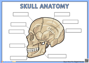
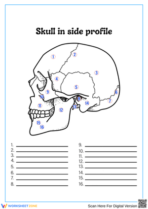
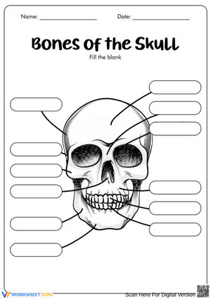
These skull anatomy worksheets printable give science teachers exactly what the topic demands: accurate multi-view diagrams, structured labeling sequences, and identification activities that move from the broadest divisions of the skull down to individual bones and the sutures that bind them. The set covers anterior and lateral perspectives — which is precisely where most anatomy instruction gets complicated — along with exercises that push students beyond recognition into genuine structural reasoning.
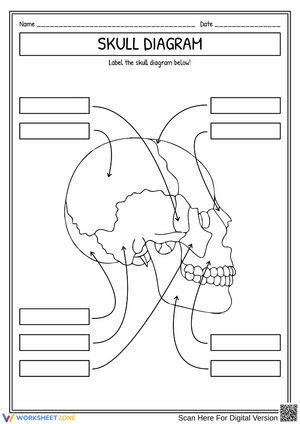
What the Worksheets Cover
Each worksheet targets a specific slice of skull anatomy rather than crowding every structure onto a single diagram. The full set addresses:
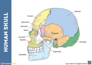
- The eight cranial bones — frontal, parietal (paired), temporal (paired), occipital, sphenoid, and ethmoid, organized by position within the protective vault rather than alphabetically
- The fourteen facial bones — mandible, maxilla (paired), zygomatic (paired), nasal (paired), lacrimal (paired), palatine (paired), inferior nasal conchae (paired), and vomer, grouped by region
- Major cranial sutures — coronal, sagittal, lambdoid, and squamosal, located precisely on both anterior and lateral diagrams
- Key anatomical landmarks — orbital cavities, nasal cavity, external auditory meatus, mastoid process, and foramen magnum, giving students the reference points that connect bone names to bone function
The cranial-versus-facial distinction anchors the entire set. Students who internalize that grouping first — eight bones forming a protective vault, fourteen forming the structural face — carry a cognitive framework that makes individual names stick far more reliably than rote list memorization ever does.
Frequent Student Errors Worth Watching For and Correcting
The sphenoid bone produces more labeling errors than any other structure in the unit. On an anterior diagram, it appears as a narrow wedge at the lateral edge of each orbit, easily overlooked between the more prominent frontal and temporal bones. On a lateral view, that same bone reads as a broad, butterfly-shaped structure with visible wing processes — a completely different visual profile. Students who label it correctly on the anterior worksheet will skip right past it on the lateral one, not recognizing it as the same bone. Presenting both views side-by-side during direct instruction, rather than on separate class days, closes that gap more reliably than any re-teaching session after the fact.
Suture names generate a second category of confusion. Coronal and sagittal are anatomical directional terms students are learning simultaneously in other contexts — a student reading "coronal suture" often retrieves "coronal plane" first, which makes the term feel redundant rather than specific. A brief explicit connection — the coronal suture runs along the coronal plane, dividing the frontal bone from the parietal bones — converts that retrieval interference into a memory hook. It takes thirty seconds and prevents the error from becoming entrenched.
A third consistent pattern: students correctly label the maxilla on the anterior diagram but cannot find it in the lateral view, where it recedes behind the zygomatic arch and looks like a background structure. These skull anatomy worksheets printable address this directly by tracing the same bone through multiple perspectives, so students encounter each structure in the visual context that actually shows up on assessments.
How to Sequence These Worksheets Across Your Unit
The most effective entry point is the anterior view — not because it is simpler, but because it maps to what students can physically locate. On the first day, before distributing anything, have students palpate their own frontal bone, zygomatic arch, nasal bones, and mandible. That tactile reference turns the diagram from abstract lines into a map of something already familiar. Students who have felt the zygomatic arch on their own face learn its name in a single exposure; students who encounter it only as a printed line take several repetitions to hold it.
The lateral view worksheet belongs in the second session, not the first. After students have anchored the major facial bones through the anterior diagram, the lateral worksheet adds the cranial vault — parietal and temporal bones, the occipital region, and the suture network — without demanding that every structure be learned at once. Introducing skull anatomy worksheets printable in this sequence follows the gradual release model: whole-class practice on the anterior view, guided work on the lateral view, then independent labeling of both for the summative check.
For five-minute bell work or exit checks, isolate a single region — just the orbital structures, or just the cranial sutures — rather than presenting a full blank skull. A partial diagram generates more precise formative data and avoids overwhelming students who are still consolidating the previous lesson. Station rotations work well at the midpoint of the unit: one station with a three-dimensional skull model, one with the printed diagrams, and students cross-reference between them. That physical-to-diagram transfer is where retention actually solidifies.
Standard Alignment
Skull anatomy instruction maps directly to the NGSS crosscutting concept of Structure and Function, which runs through both middle and high school life science. At the middle school level, MS-LS1-3 asks students to use argument supported by evidence for how the body is a system of interacting subsystems. Labeling the cranium and facial bones is the concrete, observational groundwork that makes that argument possible — students can point to the sutures between parietal and frontal bones as evidence that the skull is not a single monolithic structure but a system of interlocking parts, each with a traceable function. At the high school level, HS-LS1-2 requires students to develop and use models to illustrate hierarchical organization in multicellular organisms. Skull diagrams are exactly that kind of model. A student who identifies the mandible, traces the temporomandibular joint, and explains how its condylar process permits lateral jaw movement has met HS-LS1-2 at the organ-system level — not just labeled a picture.
Adjusting the Worksheets Across Student Readiness Levels
The most practical lever is the word bank. Adding a complete list of bone names reduces the task from pure retrieval to recognition — a meaningfully lower cognitive demand that allows students who are still building vocabulary to participate in the labeling activity rather than sit stuck with a blank page. Removing the word bank entirely, or providing a partial one that deliberately omits four or five entries, raises the demand back toward independent recall. For students who need additional reference points, a partially pre-labeled diagram — five or six bones filled in to anchor spatial orientation — lets them complete the rest without the full structure being given away.
Students working ahead of grade level benefit from the addition of specific foramina and bony processes: the foramen magnum, styloid and mastoid processes, the external auditory meatus, the mental foramen of the mandible, and the infraorbital foramen. These structures rarely appear on middle school assessments but show up consistently in high school anatomy, AP Biology, and health sciences coursework. A teacher-made overlay listing only these advanced landmarks, used alongside the standard worksheet, lets one diagram serve two instructional levels without creating a parallel document from scratch. That is worth noting for any teacher managing a heterogeneous class.
Frequently Asked Questions
How many bones does the adult human skull contain?
The adult skull contains twenty-two bones: eight cranial and fourteen facial. Some anatomical references add the three ossicles inside each middle ear and the hyoid bone in the throat, but the standard instructional count for K-12 science is twenty-two. A useful developmental connection for students: fetal skulls contain more unfused bones, and the open fontanelles present at birth are the same coronal and sagittal suture sites students label on these worksheets — a detail that tends to land well with students who have younger siblings.
What distinguishes cranial bones from facial bones?
The eight cranial bones — frontal, parietal, temporal, occipital, sphenoid, and ethmoid — form the protective vault enclosing the brain. The fourteen facial bones provide the bony structure of the face, support the eyes, nose, and mouth, and serve as attachment points for the muscles controlling expression and chewing. Teaching the functional distinction, not just the anatomical location, gives students a reasoning frame: if a bone primarily protects neural tissue, it is cranial; if it primarily supports sensory or digestive structures, it is facial.
Are these worksheets usable at both middle school and high school?
These skull anatomy worksheets printable work across both levels because the same diagram supports different task demands. Middle school teachers typically pair the anterior view with a complete word bank for first-exposure labeling. High school anatomy teachers use the identical diagram without a word bank, add suture identification, and extend into foramina and bony processes for students in advanced courses. The diagram stays constant; the task expectations around it change with the level.
How can these worksheets function as assessments?
A blank labeling diagram with numbered leader lines and no word bank produces clean, easy-to-score formative quiz data. A fifteen-item skull diagram, each structure worth one point, generates reliable evidence of recall without any grading ambiguity. For summative use, pairing a blank diagram with two or three short-answer questions about bone function — why does the occipital bone contain the foramen magnum, or what structural feature allows the mandible to move while the rest of the skull does not — pushes the assessment beyond identification into the structure-function reasoning that most state frameworks require at the secondary level.
Clear All