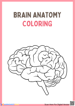
These brain printable worksheets give grades 4–8 science teachers a targeted set of exercises covering the major structures, functions, and communication pathways of the human nervous system. Each worksheet addresses a distinct anatomical concept — lobe identification, neuron structure, involuntary versus voluntary control — so teachers can assign them to fit exactly where a lesson falls within a unit rather than working through the set in one sitting. The diagrams are annotatable without confusion, and the exercises move students deliberately from identification toward application.
The Structures and Concepts Each Worksheet Covers
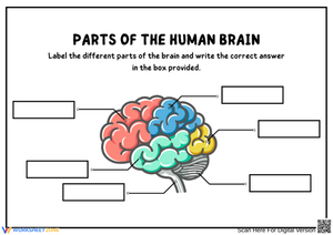
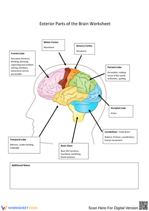
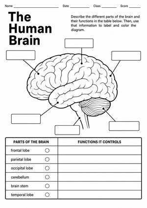
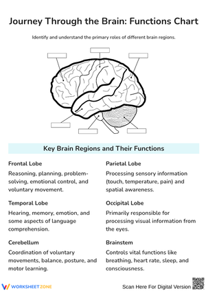
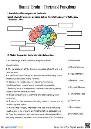
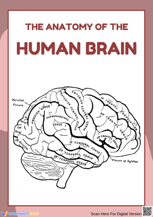
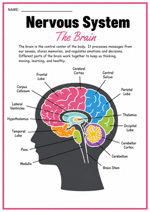
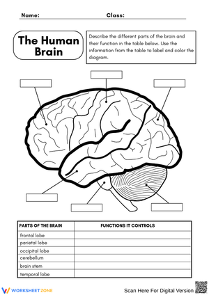
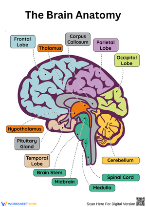
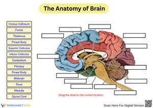
The set moves through the brain's major regions in the sequence most life science units follow. The cerebrum comes first: students label the four lobes on a lateral diagram and record each lobe's primary function in their own words. The cerebellum and brainstem each receive dedicated worksheet treatment rather than being collapsed into a generic "other structures" category — which is exactly where most textbook diagrams fall short, and a key reason students routinely conflate the two. A separate exercise covers neuron anatomy at the cellular level, walking students through signal transmission from initial stimulus to motor response.
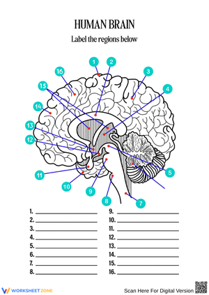
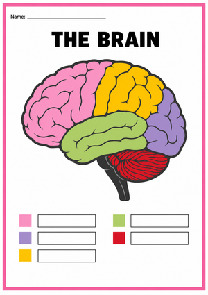
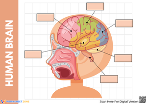
- Cerebrum labeling: students mark the frontal, parietal, temporal, and occipital lobes on a lateral diagram and note each lobe's primary function
- Lobe-to-function matching: pairs everyday scenarios — catching a ball, processing a song lyric, recognizing a familiar face — with the lobe most responsible
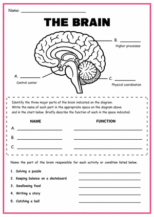
- Cerebellum and brainstem: students sort twelve bodily functions into voluntary or involuntary categories and identify the controlling structure for each
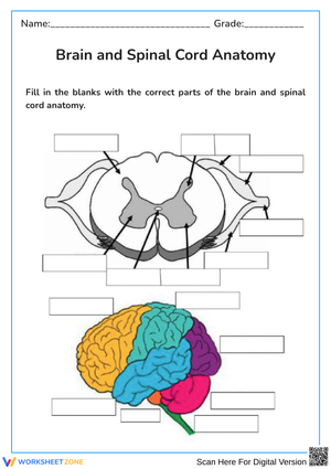
- Neuron anatomy: students label the cell body, dendrites, axon, myelin sheath, and axon terminals on a magnified diagram, then answer questions about signal direction
- Sensory pathway sequencing: students arrange stimulus-to-response steps in order and identify which neuron type — sensory, interneuron, or motor — carries each stage of the signal
Errors Students Make Consistently — and Why
The cerebrum/cerebellum confusion is the first error to anticipate. Both names begin with "cerebr-," and when students encounter them in rapid succession during a lesson, the two blur together. The most reliable fix is assigning the cerebellum a concrete, repeatable image — a tightrope walker, a surgeon holding a scalpel perfectly still — and returning to that exact image every time the structure appears in discussion or on a diagram. Once the visual anchor is in place, the name follows reliably.
Neuron signal direction is a separate, equally predictable source of confusion. Students grasp that signals travel, but a significant portion reverse the path — drawing signals entering at the axon terminal and exiting through the dendrites. A three-minute arrow exercise, done before students write anything on the neuron diagram, catches this early and takes almost no instructional time to correct. The brainstem is also consistently underestimated: students who associate complex thought with the frontal lobe conclude that more complex equals more important, mentally ranking the brainstem last. The question "which structure keeps you alive during dreamless sleep?" resets that ranking without a lecture.
Fitting These Worksheets Into a Two-Week Unit
The most productive approach is spacing the exercises across two weeks rather than assigning them back-to-back. Start with the cerebrum labeling worksheet as a pre-assessment — give it cold, before any direct instruction, and collect it. That first pass immediately shows which students arrive with prior knowledge of lobe names and which are starting from zero. Keep those early attempts and return them at the unit's end; the before-and-after comparison is one of the cleaner demonstrations of growth you can hand a student, and it costs nothing extra in planning time.
The lobe-to-function matching exercise works best as a 10-minute warm-up two or three days into instruction — after students have heard a direct lesson on the cerebrum but before they've had time to consolidate it. That specific instructional window, where information is present but not yet anchored, is where structured matching does its most useful cognitive work. The brain printable worksheets covering neuron anatomy and sensory pathway sequencing are better placed toward the end of the unit, when students have enough vocabulary to describe what they're labeling rather than simply copying terms from a reference diagram.
Adjusting the Exercises for a Range of Learners
Students who need more support get more out of the labeling exercises when you add a word bank at the bottom of the page. Six structure names printed below the diagram reduce the retrieval demand enough that students can focus on placement and function rather than spelling from memory. For those students, sequencing the matching worksheet before the blank diagram also helps — matching is lower-stakes, builds familiarity with the terms, and makes free-recall labeling feel less like a cold test.
Advanced students who finish quickly benefit from being pushed past identification into explanation. Ask them to annotate the neuron diagram with a written description of what happens chemically at the synapse — specifically the role neurotransmitters play in crossing the synaptic gap. The sensory pathway exercise becomes a genuine extension task when you remove the provided steps and ask students to write the sequence from scratch for a different stimulus: what happens, neuron by neuron, when someone hears an unexpected loud noise? That kind of generation task is where brain printable worksheets function most effectively for students who have already internalized the basic structure.
Frequently Asked Questions
What grade levels are these worksheets best suited for?
The cerebrum labeling and lobe-matching exercises work well for grades 4–6, where life science units introduce major body systems. The neuron anatomy and sensory pathway exercises are better matched to grades 6–8, where instruction expects students to operate at the cellular level and use precise anatomical terminology rather than general descriptors.
Can students complete these without a separate textbook?
The labeling and matching exercises include enough contextual information — function descriptions, structured prompts, or reference diagrams — that most students complete them without needing a separate text. The neuron pathway worksheet is the exception; students benefit from a brief direct lesson on electrical impulse transmission before attempting it independently, because the sequencing task assumes familiarity with terms like dendrite, axon, and synapse.
How much class time does each worksheet require?
Labeling and matching exercises typically run 15–20 minutes. The neuron anatomy and pathway sequencing worksheets take closer to 25–30 minutes for independent work. Budget additional time the first time students encounter a blank brain diagram — most underestimate how long accurate placement of six to eight labels takes when the image is genuinely unfamiliar, and rushing that first attempt tends to cement incorrect placements.
Do these formats match what students see on standardized assessments?
State life science assessments for grades 5–8 regularly include questions on major brain regions and nervous system function. The lobe identification and function-matching formats in these brain printable worksheets closely mirror the multiple-choice and short-answer structures students encounter on those assessments, so the practice carries direct transfer value without requiring any format adjustment on your end.