Eye Worksheets PDF: Printable Activities for Teaching Human Eye Anatomy
These eye worksheets pdf give science teachers a ready-to-print set of activities that move students from rote memorization of anatomy terms toward a working understanding of how the eye converts incoming light into something the brain can interpret as an image. The set covers both structural labeling and functional sequencing, so students build vocabulary and process knowledge at the same time. Each worksheet stands alone, meaning teachers can drop one into a warm-up block, a lab day review, or a formative check without redesigning an entire lesson plan.
Structures and Concepts Each Worksheet Addresses
The labeling worksheets in a good eye worksheets pdf set focus on the anatomical structures students will need to identify by name, location, and function. Rather than open matching, the diagrams use numbered leader lines on a clean cross-section illustration, which forces students to attend to spatial position — not just term recognition.
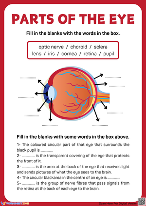
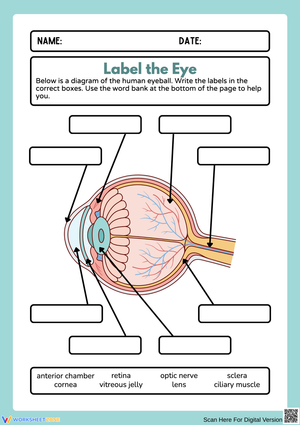
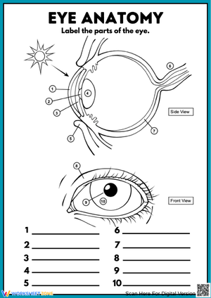
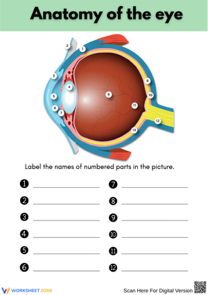
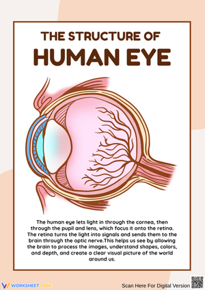
- Cornea — the transparent outer surface where incoming light first bends
- Iris — the ring of muscle that adjusts the diameter of the pupil
- Pupil — the opening that regulates how much light enters the interior
- Lens — the crystalline structure that fine-focuses light precisely onto the retina
- Retina — the light-sensitive lining containing photoreceptor cells (rods and cones)
- Optic nerve — the fiber bundle that carries electrical signals to the brain
- Sclera — the tough white outer coat that protects the eye's internal structures
Sequencing activities trace the full light pathway from cornea to occipital lobe, while a T-chart worksheet separates rod function (low-light intensity and peripheral detection) from cone function (color discrimination and fine detail). One worksheet in the set addresses vision correction, connecting myopia and hyperopia to the lens geometries used to treat them — a natural bridge to optics content in a concurrent physical science course.
The Case for Diagram-First Instruction in Eye Anatomy
Anatomy instruction benefits from what cognitive scientists call dual coding — pairing a labeled diagram with written explanation activates both visual-spatial and verbal memory pathways simultaneously, producing stronger retention than text alone. The eye is a particularly strong candidate for this approach because its structures are spatially defined and consistent: the iris sits in front of the lens, the retina lines the back interior, the optic nerve exits at a fixed point called the blind spot. Students who locate a structure on a diagram before reading about its function hold that function longer than students who encounter a glossary term first. This is the instructional logic behind diagram-first, text-second sequencing for eye anatomy units. A clean line drawing also outperforms a photograph for classroom work — photographs of human eyes rarely reveal internal layers with any useful clarity, while a well-drawn cross-section makes every structure accessible and unambiguous at a glance.
Where Students Consistently Get the Anatomy Wrong
The most reliable error pattern involves the iris and the pupil. Students understand conceptually that the pupil is an "opening," but many represent it on paper as a solid black disc — a physical membrane — rather than the absence of tissue. On labeling worksheets, this surfaces in a specific way: students swap the two labels or assign both functions to the same structure, writing "controls light entry" as the pupil's role and "the colored part" as the iris's role while attaching both descriptions to a single leader line.
A second predictable error appears on sequencing tasks. Students reliably list the correct structures in order but describe the retina as "sending an image to the brain." The retina does not transmit an image — it converts light into electrical nerve impulses, and the brain constructs the image from those signals. The fill-in-the-blank sequencing format makes this distinction visible in real time: the prompt forces students to write "electrical signals" or "nerve impulses" rather than "picture," which exposes the misconception directly in written form before it hardens into a persistent misunderstanding.
Recommended Ways to Work This Set Into Your Lesson Planning
The labeling worksheet works best before instruction, not after. Handing students a blank cross-section diagram at the start of class and asking them to fill in whatever they already know takes about four minutes and immediately surfaces vocabulary gaps. During direct instruction, students mark corrections in a different color on the same sheet — growth becomes visible without requiring a second document. The sequencing worksheet fits naturally at the end of a lesson as a formative check: if students can trace the light pathway without consulting notes, the concept has moved from short-term exposure toward durable understanding. The rods-and-cones T-chart fits a station-rotation model well — one station for the worksheet, one for a brief reading excerpt from the National Eye Institute, one for a short flashlight-and-colored-filters demonstration. Rotating groups every eight to ten minutes keeps the period moving without fragmenting the content.
One pairing worth building into the unit: run the labeling worksheet immediately before a preserved eye dissection. When students have just written "lens" on a diagram and then hold a cow-eye lens and observe its convex shape and transparency firsthand, that label stops being a vocabulary item and becomes a real object they have examined. The same eye worksheets pdf set works as a strong post-dissection check — students revisit the labeled diagram and annotate each structure with one direct observation from the dissection, connecting the printed illustration to the physical specimen they just handled.
Standard Alignment
These worksheets align to NGSS MS-LS1-8, which addresses how sensory receptor cells respond to stimuli by sending signals that are processed by the brain. The sequencing worksheet directly targets this standard: students must articulate the complete chain from physical stimulus (light hitting the cornea) to neural signal (impulses traveling along the optic nerve) to brain interpretation (occipital lobe processing). MS-LS1-8 requires students to describe a cause-and-effect mechanism — not simply name anatomical parts — and the sequencing format demands exactly that kind of explanatory writing. For high school courses, the vision correction worksheet connects to HS-PS4-5, which involves using models to explain how electromagnetic radiation, including visible light, behaves when passing through lenses of different geometries.
Adjusting the Set for a Range of Learners
For students working below grade level, attach a completed reference diagram — a small, fully labeled version of the same cross-section — to the bottom of the labeling worksheet. This allows students to self-check structure by structure rather than stalling at the first blank and disengaging. A word bank that pairs each term with a brief definition (not just the term alone) serves the same purpose for students who have a vocabulary gap rather than a processing gap. English learners benefit from bilingual label options or a glossary sidebar; the diagram format is inherently accessible because spatial relationships carry meaning even when individual terms are unfamiliar.
Students who finish the standard worksheet quickly need a harder prompt, not a second copy of the same task. Remove the word bank entirely and add an open-response extension: "The fovea is the cone-dense center of the retina. Predict how your vision would change if the fovea were damaged, and explain your reasoning using what you know about rod and cone function." That question requires students to synthesize photoreceptor function with spatial anatomy — moving well past label recall into genuine application thinking.
Frequently Asked Questions
Do these worksheets include an answer key?
Each worksheet in the set comes with a corresponding answer key. The labeled diagram key includes correct structure placement, and the sequencing key shows the complete light-pathway chain with accepted phrasing for the nerve-impulse steps. The rods-and-cones T-chart key includes sample responses for any open-ended comparison items, giving teachers a consistent grading reference across class sections.
What grade levels fit this material best?
The labeling and sequencing worksheets are written for grades 6–8, where eye anatomy appears most frequently in life science units. The vision correction worksheet, which introduces refraction and lens geometry, is better suited to grades 9–10 — in either a biology course or a physics course covering optics. The differentiation adjustments described above extend usability across this full range without requiring teachers to build entirely separate materials for different sections.
Can a worksheet replace direct instruction on the eye?
The eye worksheets pdf set reinforces and assesses understanding — it does not introduce content cold. A student who encounters "optic nerve" for the first time on a blank labeling diagram without any prior instruction will guess, not learn. The intended workflow: use the labeling worksheet as a pre-assessment before instruction begins, then use the sequencing worksheet as a formative check after the lesson. That bracketing structure makes each worksheet purposeful rather than merely supplementary.
Are the diagrams sharp enough to display under a document camera?
Yes. The cross-section illustrations use clean line work with enough white space that projecting under a document camera or displaying a scanned copy on a classroom screen works without loss of detail. Teachers who model the labeling process by completing the worksheet on screen while students follow along on their printed copies report that the walk-through catches sclera-cornea confusion early — students frequently misplace both structures on the outer surface of the diagram, and seeing the correct placement projected in real time closes that gap before it shows up on an assessment.
Clear All