These quad muscle printable pdf worksheets for 10th grade give anatomy and biology teachers a set of labeling and analysis activities organized around the quadriceps femoris — the four-muscle group on the anterior thigh that students need to understand structurally before they can reason about knee mechanics, injury patterns, or neural innervation. Each worksheet targets a distinct piece of that content, so teachers can pull one into a lesson without reorganizing the whole unit.
What Each Worksheet Covers
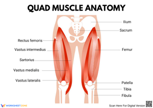
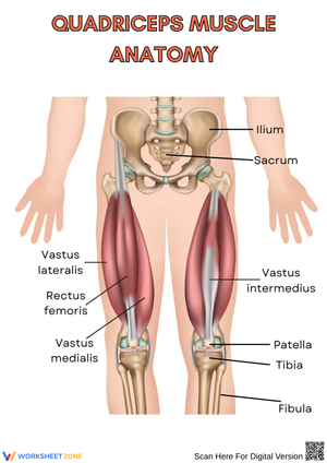
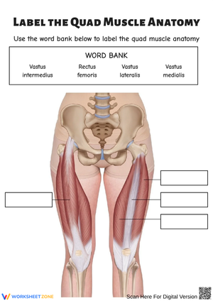
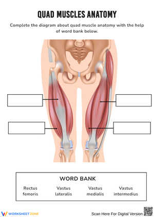
The set works through quadriceps anatomy in a logical order. Labeling worksheets come first: students identify all four muscle heads on an unlabeled anterior thigh diagram — rectus femoris, vastus lateralis, vastus medialis, and vastus intermedius — then annotate each with its position relative to the others. Once names and spatial positions are established, later worksheets shift to origins and insertions. Students mark the anterior inferior iliac spine as the rectus femoris origin and identify the separate femoral origins of the three vasti, which sets up the explanation of why the rectus femoris is the only bi-articular muscle in the group, capable of acting at both the hip and the knee.
The remaining worksheets in the set address:
- The full insertion chain — from the quadriceps tendon through the patella and patellar ligament to the tibial tuberosity — requiring students to label each segment separately
- The agonist/antagonist relationship between the quadriceps and hamstrings during knee extension and flexion
- The femoral nerve pathway from the L2–L4 lumbar segments through the femoral triangle to the anterior thigh musculature
- A patellar reflex diagram, where students annotate the sensory and motor limbs of the reflex arc and identify the spinal level involved
Student Mistakes Worth Catching Early
The most persistent error in student diagrams on this topic is misplacing the vastus medialis. Students know it sits on the medial side of the thigh, so they draw it running the full length of the inner thigh — but the defining oblique fibers are concentrated in the distal third, close to the knee. A student who draws the vastus medialis as a structural mirror of the vastus lateralis has missed the anatomical point entirely, and that error rarely becomes visible until a labeled-diagram quiz reveals it.
The quadriceps tendon and patellar ligament distinction creates a second consistent problem. Students frequently write that the quadriceps attaches directly to the tibial tuberosity, skipping the patella entirely. Others call the patellar ligament a tendon — understandable, since the structure feels continuous — but the segment below the patella is technically a ligament because it connects bone to bone. These worksheets require students to label every segment of that insertion chain separately, which forces the distinction into the activity rather than leaving it to last-minute review.
A third sticking point: under time pressure, students write "femur" as the origin for all four quadriceps muscles. The rectus femoris originates at the anterior inferior iliac spine on the pelvis, and that pelvic attachment is precisely what allows it to flex the hip as well as extend the knee. The labeling activities flag that origin point explicitly so it doesn't slip past students before the unit test.
Fitting These Worksheets Into Your Weekly Plan
The labeling worksheets work best as the first activity after introducing the quadriceps — typically 15 to 20 minutes of a 50-minute class period, leaving time for whole-class comparison of placements. A brief kinesthetic check before the worksheet helps: ask students to stand and perform one slow bodyweight squat with a hand on the front of the thigh just above the knee. Most can feel the rectus femoris tighten on the way down, and that physical reference consistently improves retention of the muscle's position and function when they return to the diagram.
The origin/insertion worksheet pairs well with a projected diagram that the class traces together before students mark the same attachment points on their own worksheet. The femoral nerve mapping worksheet fits mid-unit once spinal plexus organization has been introduced, but it also runs cleanly in the last 10 minutes of class as a memory-retrieval check — students trace the nerve pathway without prompts, then compare to a key. That quick exercise functions as a clean formative read before a written assessment. The quad muscle printable pdf worksheets for 10th grade work across all of those placement options because each stands alone, without requiring the previous worksheet to be completed first.
Adapting the Worksheets for a Range of Learners
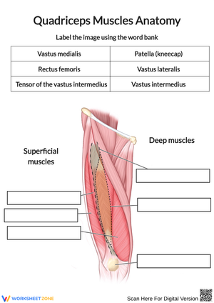
Students who need additional support work from a diagram with the muscles pre-colored by anatomical layer — superficial muscles in one color, the deep layer in another. They add labels to regions that are already visually distinguished, which removes one layer of spatial reasoning (particularly useful for the vastus intermedius, which is invisible in a standard surface view) while still holding students accountable for names, positions, and functions.
Students working ahead receive the blank-canvas version: an outline of the thigh with no muscle shapes drawn in, requiring them to reconstruct approximate muscle positions from memory before labeling. That demands a different kind of spatial recall than pointing to a pre-drawn shape. For the strongest students in a course combining anatomy and physiology, a brief extension asks them to treat the knee as a simple hinge and estimate — using basic torque ratios, no calculus required — how much force the quadriceps tendon must generate to hold the leg extended against resistance at the ankle. The arithmetic is accessible at 10th grade, and the cross-disciplinary thinking is exactly what distinguishes advanced work in this unit. These quad muscle printable pdf worksheets for 10th grade include both the pre-colored and blank-canvas diagram versions, so teachers don't need to build a separate resource for differentiation.
Standard Alignment
NGSS HS-LS1-2 (From Molecules to Organisms: Structures and Processes) asks students to develop and use models illustrating how systems of organs work together to support whole-organism function. The labeling and tracing activities in this set address that standard directly: students model the anterior thigh as an integrated system in which four distinct muscles share a tendon, incorporate a sesamoid bone, and transmit force through a ligament to act on the tibia. The femoral nerve worksheet extends the standard into neural coordination, connecting the spinal segments responsible for innervation to the muscular response — with the patellar reflex as the concrete teaching example.
State anatomy and physiology frameworks at the 10th grade level typically expect mastery of origin/insertion vocabulary, agonist/antagonist relationships, and peripheral nerve organization. The quad muscle printable pdf worksheets for 10th grade address all three and also align with the reflex arc content common in health science and sports medicine pathways, since the patellar reflex diagram traces the full L4 arc from stretch receptor activation to motor response.
Frequently Asked Questions
Do these worksheets require prior anatomy background, or can they be used at the very start of the quadriceps unit?
The labeling worksheets work at the start of instruction — students can treat them as first contact with the material, filling in what they recognize and leaving blank what they don't yet know. The origin/insertion and nerve mapping worksheets assume familiarity with vocabulary like origin, insertion, and spinal segment, so those fit better mid-unit or as review activities. Because each worksheet stands alone, teachers can pull whichever piece fits the day's lesson without working through the set in sequence.
Are the diagrams detailed enough to show the vastus intermedius?
Because the vastus intermedius is entirely hidden beneath the rectus femoris in a standard surface view, the set includes a layered diagram that removes the superficial muscles to expose the deep layer. Students complete the full surface-view diagram first, then work from the layered version to add the vastus intermedius. That two-step sequence pushes students to think about anatomical depth — exactly the kind of spatial reasoning that practical anatomy assessments target.
Is the femoral nerve worksheet appropriate for a standard biology class, or is it better suited to an anatomy elective?
The nerve mapping worksheet works in both settings. In a general biology class, it introduces peripheral nervous system structure through a concrete example most students have already encountered — the patellar reflex at a routine medical appointment. In an anatomy elective, it goes further: students trace the lumbar plexus origin at L2–L4, mark the femoral triangle landmarks, and distinguish motor branches from sensory branches. Both levels of the worksheet are included in the set, so teachers assign the version that matches their course.