Foot Muscle Worksheets PDF for 10th Grade
These foot muscle worksheets pdf for 10th grade move students past the general body-systems survey that fills most of the first semester and into the functional specifics of the lower limb — how muscles are classified by their points of origin, how they stack in layered arrangements under the sole, and what that structure actually accomplishes during walking, jumping, and balance recovery. The set gives anatomy teachers standalone worksheets that drop into an existing unit as formative checks after a model lab, as structured review before a practical exam, or as targeted practice after direct instruction on the plantar layers.
Muscle Groups and Structures Addressed in Each Worksheet
The organizing framework throughout the set is the intrinsic/extrinsic classification. Extrinsic muscles — the gastrocnemius, soleus, tibialis anterior, fibularis longus — originate in the leg and drive the larger, power-oriented movements of the foot: plantarflexion, dorsiflexion, inversion, eversion. Intrinsic muscles have both their origin and insertion below the ankle joint and handle fine toe control and dynamic arch stabilization. Students work through both groups before the worksheets move into layer-by-layer identification of the plantar surface.
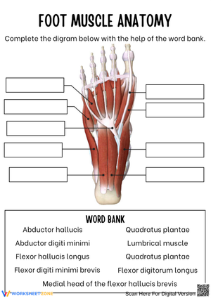
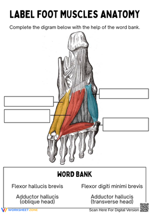
The plantar layers get the most concentrated treatment. Each worksheet moves through all four layers from superficial to deep. The first layer holds three muscles — abductor hallucis along the medial edge, flexor digitorum brevis in the center, and abductor digiti minimi on the lateral side. The second layer introduces the quadratus plantae and the four lumbrical muscles, with specific attention to how the quadratus plantae redirects the diagonal pull of the flexor digitorum longus tendon so the toes flex in a straight line rather than at an angle. The third layer addresses the flexor hallucis brevis, adductor hallucis, and flexor digiti minimi brevis; the adductor hallucis gets extended treatment because its oblique and transverse heads form a configuration students often describe as a "7," which turns out to be a useful visual anchor. The fourth layer covers the plantar and dorsal interossei, situated between the metatarsal bones and responsible for toe adduction and abduction respectively.
Biomechanical function runs alongside identification throughout. The windlass mechanism — the increase in plantar fascia tension when the big toe is extended — appears in several worksheets as an applied reasoning question rather than a vocabulary term to copy down. The tibialis posterior and fibularis longus are presented as the stirrup-like force couple that elevates the arch dynamically, not merely as named structures to locate on a diagram. The plantar layer content in a foot muscle worksheets pdf for 10th grade set that covers all four layers tends to be the most time-intensive material students encounter in a lower-limb anatomy unit — and the most assessable.
Student Errors Worth Anticipating Before You Hand These Out
The most consistent error on intrinsic/extrinsic classification tasks: students decide a muscle is intrinsic based on what it does rather than where it originates. They know the gastrocnemius moves the foot, so they categorize it as a foot muscle. One sentence clears this up — intrinsic means the muscle belly lives entirely below the ankle, not that it acts on the foot — but it needs to land before the first identification exercise, not after a stack of wrong answers.
The quadratus plantae reliably confuses students who have internalized that muscles insert into bone. When they read "inserts into the tendon of flexor digitorum longus," they treat it as an exception to memorize rather than a structural solution to a mechanical problem. Once they understand that the FDL approaches the foot at a diagonal and the quadratus plantae corrects that angle so the toes flex straight, the anatomy stops feeling arbitrary — and the detail stays in memory.
Layer ordering on diagram tasks is a third sticking point. Students working from a flat illustration frequently swap the second and third plantar layers because they haven't yet built a three-dimensional mental picture of the sole. Color-coding by layer on the first pass through a worksheet, then asking students to reproduce the same diagram from memory without color support on a subsequent attempt, addresses this before it becomes a persistent confusion.
Building These Worksheets Into Your Anatomy Unit
Sequence the extrinsic muscle worksheets before the plantar layer materials. Students who don't yet have a clear picture of where the leg musculature ends and the foot begins will struggle with intrinsic classification — the distinction only carries meaning once the extrinsic side is solid.
Before any plantar identification worksheet, run a short kinesthetic moment: ask students to remove their shoes and try to pull the ball of the foot toward the heel without curling the toes. They feel the abductor hallucis fire. That 90-second physical experience does more to anchor "intrinsic muscle function" than several minutes of note-copying. Similarly, asking students to pull their own big toe upward and notice the change in tension along the sole demonstrates the windlass mechanism better than any diagram. When a foot muscle worksheets pdf for 10th grade is paired with this kind of brief physical prompt before students sit down with the diagram, retention at the one-week check is noticeably stronger.
Sports-injury connections are worth building in deliberately. Many 10th graders are active athletes, and mapping familiar injury names onto the anatomy they are studying converts abstract content into something personally relevant. Turf toe involves the flexor hallucis brevis; plantar fasciitis implicates the first-layer intrinsics and the plantar fascia together; shin splints draw in the extrinsic anterior compartment muscles. These aren't tangential examples — they're the same structures on the worksheet, viewed from a clinical angle, and they consistently raise engagement among students who otherwise find muscle memorization tedious.
Adjusting for Different Student Levels Within the Same Unit
For students who need more direct support, limit the first round of worksheets to the first plantar layer and two or three extrinsic muscles most relevant to walking. Trying to hold all four layers at once before the first layer is solid reliably produces copying behavior rather than actual reasoning. Work layer by layer with brief verbal checkpoints — not full quizzes — before moving deeper.
For students ready to extend, add a gait cycle application to each identification worksheet: mark which phase — heel strike, mid-stance, push-off — each muscle is most active during, and explain why. This moves the task from passive labeling to functional analysis and is a considerably more appropriate challenge for students headed toward AP Biology, sports medicine, or any kinesiology pathway. The step up in cognitive demand is real, and students who need that level of challenge recognize it immediately.
Standard Alignment
NGSS HS-LS1-2 asks students to develop and use models to illustrate how structure enables function in organisms. The foot's four-layer plantar arrangement, its arch mechanics, and the interplay between intrinsic and extrinsic muscle groups give students a direct, concrete line from structural organization to functional outcome — exactly what the standard targets. In state-level anatomy and physiology frameworks used in California, Texas, and Florida Health Science pathways, 10th graders are expected to identify major muscle groups of the appendicular skeleton and describe their roles in movement. The plantar layers address that expectation at a specific and assessable level of detail rather than at the level of general vocabulary.
Frequently Asked Questions
What distinguishes intrinsic foot muscles from extrinsic ones?
Intrinsic muscles have both their origin and insertion within the foot, below the ankle joint. They handle fine toe movements and dynamic arch support. Extrinsic muscles originate in the leg and enter the foot via long tendons, providing the force for larger movements — plantarflexion, dorsiflexion, inversion, and eversion. The key distinction is the location of the muscle belly, not the site of action.
How many layers are in the sole of the foot, and what does each one contain?
Four plantar layers. The first and most superficial holds the abductor hallucis, flexor digitorum brevis, and abductor digiti minimi. The second layer contains the quadratus plantae and four lumbrical muscles. The third layer includes the flexor hallucis brevis, adductor hallucis, and flexor digiti minimi brevis. The fourth and deepest layer contains the plantar and dorsal interossei, which occupy the spaces between the metatarsal bones.
Why is the quadratus plantae considered anatomically unusual?
Most muscles insert into bone. The quadratus plantae inserts instead into the tendon of the flexor digitorum longus and functions to correct the diagonal angle of that tendon's pull, keeping the toes flexing straight rather than laterally. Treating this as a functional explanation — rather than a memorized exception — is a consistent point of emphasis in any foot muscle worksheets pdf for 10th grade that covers the second plantar layer with genuine depth.
Which muscles are primarily responsible for maintaining the foot arches?
Arch support is divided between static structures — ligaments and the plantar fascia — and dynamic ones. The tibialis posterior and fibularis longus create a stirrup under the foot that actively elevates the arch during movement. Among the intrinsic muscles, the abductor hallucis and flexor digitorum brevis contribute first-layer support, while the adductor hallucis — through its oblique and transverse heads — specifically maintains the transverse arch.
Do the worksheets address the dorsal surface of the foot as well as the plantar layers?
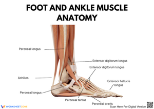
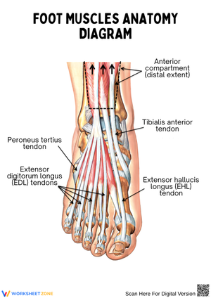
The dorsal surface has far fewer intrinsic muscles — primarily the extensor digitorum brevis and extensor hallucis brevis — and the set focuses most of its detail on the plantar layers, which are anatomically denser and more heavily assessed. That said, the dorsal interossei from the fourth plantar layer also act on the dorsal surface, so students who have worked through all four layers will encounter dorsal function within the plantar-layer worksheets themselves.
Clear All