These 10th grade deltoid muscle pdf worksheets give anatomy and biology teachers a structured set of labeled-diagram exercises, fiber function analysis tasks, and clinical application case studies built for the Grade 10 muscular system unit. The set moves students from basic structural identification — origins, insertions, the three fiber groups — through nerve pathway reasoning and real-world injection site analysis.
What Each Worksheet Covers
The deltoid works well as a focal structure at this level because it requires students to hold three anatomically distinct regions in mind simultaneously while connecting them to a single insertion point and a shared nerve supply. The 10th grade deltoid muscle pdf worksheets in this set each target a distinct layer: one worksheet addresses structural labeling, another pushes into functional analysis, a third handles nerve pathway mapping, and the final pair present clinical case scenarios.
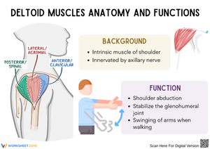
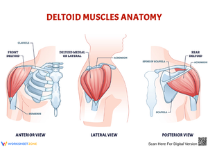
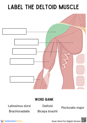
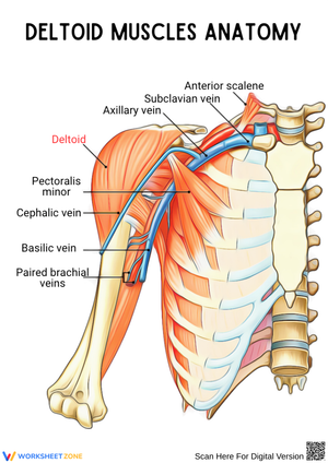
- Structural labeling: Students annotate the anterior (clavicular), lateral (acromial), and posterior (spinal) fiber groups on unlabeled shoulder diagrams, identifying origin points on the lateral third of the clavicle, the acromion, and the spine of the scapula, then marking the shared insertion at the deltoid tuberosity on the humerus.
- Functional analysis: Given a specific arm movement — throwing a baseball, reaching across a table, pulling a door open — students identify which fiber group is the prime mover and write a brief mechanical explanation connecting origin and insertion to the direction of pull.
- Nerve pathway mapping: Students trace the axillary nerve from the C5 and C6 roots through the brachial plexus to the muscle, then work through scenario questions about which shoulder movements would be lost after a fracture of the surgical neck of the humerus.
- Clinical case studies: Short patient scenarios require students to explain deltoid injection site selection, identify signs of shoulder impingement, and connect axillary nerve damage to specific functional deficits.
- Skeletal cross-reference: One worksheet sends students to a separate skeletal diagram to locate the acromion, spine of scapula, and deltoid tuberosity before returning to the muscle labeling task — reinforcing that muscular and skeletal anatomy are one integrated system, not two sequential units to be memorized in isolation.
Three Fibers, Three Distinct Jobs
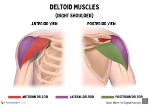
The anterior fibers — originating from the lateral third of the clavicle — handle flexion and internal rotation. The lateral fibers, arising from the acromion, are the primary drivers of abduction above 15 degrees; it is the supraspinatus that initiates that first arc of movement. The posterior fibers, anchored to the spine of the scapula, manage extension and external rotation, the motion pattern behind pulling a rowing oar or drawing the arm back before a throw.
All three sets converge at the deltoid tuberosity, the roughened V-shaped patch on the lateral shaft of the humerus. What students often find genuinely surprising is that the anterior and posterior fibers are functional antagonists — they share a muscle belly but oppose each other depending on which movement is demanded. That concept is a productive entry point into broader discussions of agonist-antagonist pairs, and it appears explicitly on the functional analysis worksheet, where students must explain how one structure can produce opposing actions.
Lesson-Planning Strategies to Get the Most From This Set
The labeling worksheet works best early in the unit, before students have any terminology memorized, because physically writing labels on an unlabeled diagram builds spatial memory more reliably than reading a textbook description. Assign it during the first lesson on shoulder anatomy, then follow with the functional analysis worksheet after the lecture on glenohumeral joint mechanics. Save the clinical case studies for the end of the unit, when students can draw simultaneously on their knowledge of origins, insertions, and nerve supply.
These 10th grade deltoid muscle pdf worksheets lend themselves to a color-coding activity that takes about 12 minutes and consistently produces better retention than an additional reading: students shade the three fiber groups in different colors, then stand and perform the movement each color represents while pressing a hand to their own shoulder. That kinesthetic step bridges the gap between a flat printed diagram and a three-dimensional movement pattern in a way that re-reading labels does not. For the clinical case studies, assigning groups of three — rather than individual silent work — surfaces the reasoning. Small groups argue through the logic aloud and catch each other's terminology errors before writing them down.
Common Student Errors Worth Catching Before the Unit Assessment
The most persistent error on the functional analysis worksheet is treating all three fiber groups as abductors. Students learn "the deltoid abducts the arm" and apply that to every fiber indiscriminately. The prompt "which fibers are most active during a military press?" exposes this reliably: students who wrote "all three equally" were applying a shortcut rather than reasoning from origin-insertion mechanics. The worksheet's fiber-specific movement questions are structured so that shortcut fails, which is precisely the point.
On the nerve pathway worksheet, students frequently place axillary nerve damage at the cervical spine rather than at the shoulder. They know C5-C6 are involved and assume the injury site is spinal — they haven't yet built a working mental model of how peripheral nerves travel distally from the plexus before reaching the target muscle. A simple clarifying question mid-class — "where along the nerve's route would a shoulder dislocation most likely cause damage?" — usually breaks the misconception. The clinical case studies reinforce this distinction, which is why they should follow the nerve pathway mapping exercise rather than precede it.
Standard Alignment
NGSS HS-LS1-2 asks students to develop and use a model to illustrate how interacting systems within multicellular organisms provide specific functions within those organisms. The labeling and functional analysis worksheets address this directly: students model how the skeletal, muscular, and nervous systems interact at a single joint to produce and constrain movement. The nerve pathway mapping worksheet adds the nervous system to that model explicitly, making all three systems visible at once. In most 10th grade sequences — including those following TEKS Biology in Texas and California's NGSS-aligned frameworks — this material falls in the human body systems unit, typically mid-year after cells and tissues have been covered.
Adjusting the Set for a Range of Learners
Students who struggle with spatial reasoning find the labeling worksheet more accessible when they have a reference image to orient from — a 3D shoulder diagram or a physical model they can rotate — before working on the flat printed version. The barrier for these students is usually not reading difficulty or terminology load; it is mentally rotating a 2D image to match a remembered orientation. Providing a reference image alongside the blank diagram shifts the cognitive demand toward functional recall rather than visual-spatial processing.
Students who move through the basic material quickly can extend into the multipennate architecture of the lateral fibers — why that pennation pattern concentrates more force-generating fibers per unit volume than a parallel fiber arrangement — and apply it to a lever-system calculation of mechanical advantage during arm abduction. That cross-curricular math extension works especially well for students who engage more readily with physics-style problem solving than with clinical narrative scenarios.
Frequently Asked Questions
Do these worksheets require a specific textbook or prior unit?
No specific textbook is required. The 10th grade deltoid muscle pdf worksheets are self-contained: each worksheet includes the reference terminology and diagram context students need to complete the tasks. A prior unit covering basic bone anatomy — specifically the clavicle, scapula, and humerus — makes the labeling worksheets more efficient, but the skeletal cross-reference worksheet in the set addresses that context directly for classes that haven't covered it yet.
How long does each worksheet take to complete in class?
The structural labeling worksheet typically runs 15 to 20 minutes depending on how much discussion the teacher builds into it. The functional analysis worksheet takes 20 to 25 minutes if students write out their mechanical reasoning rather than just circling answers. The nerve pathway mapping and clinical case study worksheets run longer — 25 to 35 minutes each — and work well as extended work or as the basis for a short group presentation activity at the end of the unit.
Are the clinical case studies accessible to standard-track students, or are they only suitable for advanced biology?
The case studies are written for standard 10th grade science vocabulary — they don't assume prior exposure to clinical terminology beyond what the earlier worksheets introduce. The axillary nerve damage scenario and the injection site reasoning exercise both require students to apply knowledge from the labeling and nerve pathway worksheets, so assigning them in sequence keeps the language accessible without removing the analytical challenge that makes them useful.