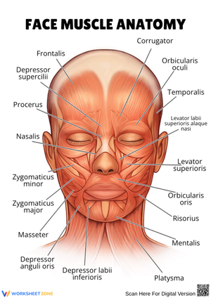
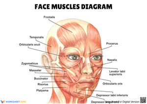
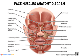
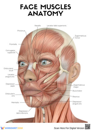
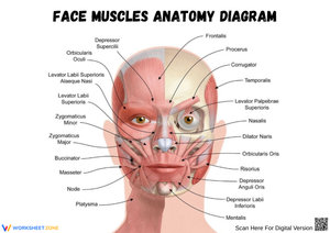
These 10th grade face muscle worksheets printable resources give biology teachers a focused set of labeling, matching, and short-answer exercises built around the muscles of facial expression and mastication. The set covers the major mimetic muscles — orbicularis oculi, zygomaticus major and minor, frontalis, buccinator, and corrugator supercilii — alongside the masticatory group: the masseter, temporalis, and pterygoids. Anterior and lateral view diagrams give students two spatial angles on a structure most of them have never consciously tried to analyze before.
The Structural Distinction That Actually Shapes This Unit
Most of the muscular system unit up to this point follows a consistent logic: muscle originates on bone, inserts on bone, crosses a joint, and moves the skeleton. Facial muscles break that rule. The muscles of facial expression typically originate from bone or fascia and insert directly into the dermis — when they contract, they pull the skin rather than rotate a joint. That difference throws students who have just internalized the standard origin-insertion model, and it needs explicit address before labeling exercises can do much good.
A second distinction worth teaching early is the two separate nerve pathways governing this region. Cranial Nerve VII, the facial nerve, drives the muscles of expression — every smile, squint, and furrowed brow. Cranial Nerve V, the trigeminal, controls the muscles of mastication, with the masseter and temporalis doing the heavy mechanical work of chewing. Students routinely conflate these two groups because they sit close together on any diagram. Keeping them visually and functionally separated from the first exercise helps the nerve distinction hold before assessment arrives.
Skills Each Worksheet Builds
The 10th grade face muscle worksheets printable materials target three types of thinking in sequence. First, spatial recall: students identify named muscles on unlabeled diagrams from both anterior and lateral views. Second, functional mapping: matching each muscle to its primary action — zygomaticus major elevates the corners of the mouth, corrugator supercilii draws the brows medially, buccinator compresses the cheek wall. Third, applied reasoning: short-answer prompts ask students to explain what would happen to a person's smile if Cranial Nerve VII were damaged on one side, or why the masseter is classified as masticatory despite its visible position on the face.
That third category is where these exercises push past standard anatomy fill-ins. Bell's Palsy works as a clinical anchor because most tenth graders can immediately picture unilateral facial drooping, and it makes the nerve distinction feel consequential rather than terminological.
Building These Worksheets Into Your Anatomy Unit
A strong entry sequence is to run the introductory labeling exercise during the lesson where you first teach the difference between mimetic and masticatory muscles. Before distributing the worksheet, give students two minutes with small hand mirrors at their desks. Ask them to contract specific muscles deliberately — raise the frontalis, close the orbicularis oculi tightly, activate the zygomaticus. Then hand out the diagram. Students who just watched their own face move label muscle locations noticeably more accurately than students who go straight to the diagram cold.
The matching and applied-reasoning exercises work well as mid-unit formative checks, roughly two days in. The blank recall diagram — no word bank — belongs at the end as a pre-test or quiz template. Using 10th grade face muscle worksheets printable resources as peer-review tools on that final pass is worth the extra setup time: students catch each other's misplaced labels on the pterygoids or the buccinator faster than a teacher scanning thirty papers at once, and the conversation that follows reliably surfaces the misconceptions worth addressing before a major exam.
Errors Students Make That These Exercises Reveal
The most consistent error is classifying the masseter as a muscle of facial expression. It sits prominently on the cheek and shows clearly on lateral diagrams, so students assume it belongs with the mimetic group. The clinical angle helps correct this: ask which cranial nerve serves the masseter — Cranial Nerve V, not VII — and the functional category usually locks in. Worksheets that use separate color coding for expression muscles and masticatory muscles make that separation visible before the error fully forms.
A second recurring problem is transposing orbicularis oris and orbicularis oculi — not just the names but the locations on the diagram. Both are circular sphincter-type muscles, which is unusual, and students who memorize one sometimes mislabel the other. Spending thirty seconds on the Latin etymology — oris for mouth, oculi for eye — gives students a retrieval cue that outlasts the unit.
A subtler error involves the zygomaticus major and minor. On most diagrams they look nearly identical in position, but they produce slightly different pulls: the major lifts the corner of the mouth outward and upward, while the minor elevates the upper lip. A worksheet exercise that asks students to trace each muscle's fiber direction and label its pull vector — rather than just write the name — makes that distinction visible before the exam rather than on it.
Standard Alignment
These worksheets align with NGSS HS-LS1-2, which asks students to use models to illustrate how the structures of organisms enable their functions. Labeled anatomical diagrams directly meet that expectation: students work with a structural representation and connect each labeled part to a biological function. The nerve pathway content also supports HS-LS1-3, which addresses information processing in biological systems — the facial nerve directing expression and the trigeminal directing mastication are a clear, observable example of how nervous system signals produce specific muscular responses.
Adjusting the Set for Different Learners
Students who need additional support do well with a partially labeled diagram — several muscles pre-filled, the rest blank — before moving to fully unlabeled exercises. Providing a printed vocabulary reference with Latin root breakdowns (orbicularis from orbis, meaning ring; zygomaticus from zygoma, the cheekbone arch) gives struggling students a decoding strategy rather than a list to memorize through repetition alone. For students who move quickly through labeling, the clinical short-answer prompts — hypothesizing which expressions a patient with Bell's Palsy would lose, or explaining why a mandibular fracture impairs chewing but not blinking — push toward analysis without requiring separate materials.
Frequently Asked Questions
Are these worksheets appropriate for students who haven't studied the nervous system yet?
Yes, with brief front-loading. The labeling and matching exercises work independently of prior nervous system knowledge. The Cranial Nerve VII versus Cranial Nerve V distinction is worth introducing as a short teacher explanation even before a full nerves unit — students need a functional reason to separate the two muscle groups rather than lumping them by location. A complete prior unit is not required.
How many class periods does this set typically require?
Plan for approximately three periods if you use the full sequence: introductory labeling with a word bank, functional matching, and blank recall. Individual worksheets stand alone as single-period activities or as ten-minute warm-up tasks spread across several days. The clinical short-answer sections sometimes run long when classes engage with the Bell's Palsy or mastication questions — worth building in buffer time if your students tend to run with application prompts.
Do these resources fit AP Biology and anatomy electives, or are they calibrated strictly for standard 10th grade biology?
The labeling and matching content fits a standard 10th grade biology course without modification. For AP Biology or a dedicated anatomy elective, the clinical short-answer sections carry the most weight — they ask for the structural-functional reasoning those courses require. The 10th grade face muscle worksheets printable resources can be paired with more detailed reference diagrams in an anatomy elective or used as introductory material before moving into the deeper musculature of the pterygoids and the mechanics of the temporomandibular joint.