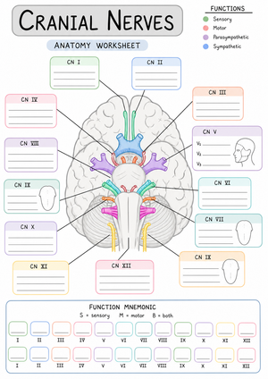
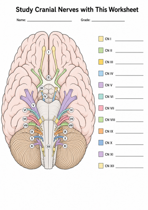
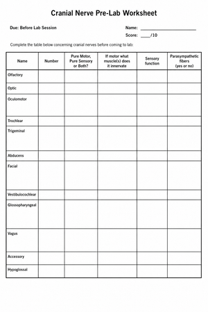
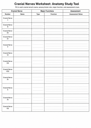
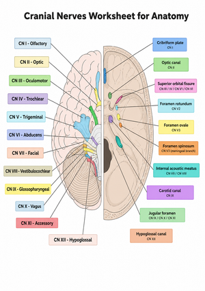
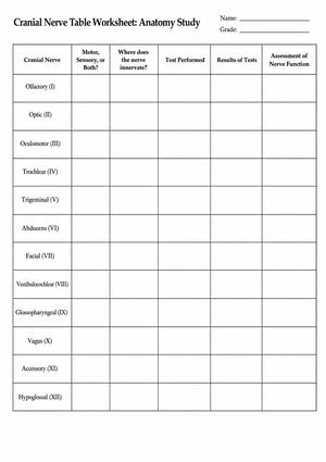
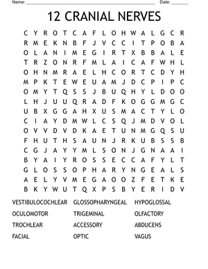
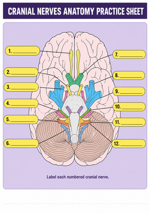
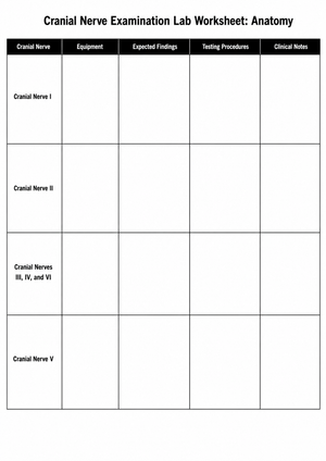
These cranial nerves printable worksheets for 11th grade give anatomy teachers a structured path through one of the most detail-heavy topics in secondary biology — twelve nerve pairs, each with a Roman numeral designation, a functional classification, a brainstem or forebrain origin, a specific skull exit point, and connections to target organs that can stretch from the eye socket to the digestive tract. The set moves from identification through functional reasoning, so students aren't just naming nerves; they're explaining why a brain-originating nerve needs to reach the stomach.
What the Set Covers
Identification comes first. Early worksheets ask students to match each nerve to its Roman numeral and classify it as sensory, motor, or mixed. That sounds manageable until a student realizes CN VIII (Vestibulocochlear) handles both hearing and balance under a single classification, or that CN V (Trigeminal) is the largest cranial nerve by diameter despite sharing the mixed category with nerves that feel far more complex on paper. Getting these classifications accurate before moving forward matters — later tasks depend on them.
From there, the worksheets shift to origin and exit points. Students label brainstem zones — midbrain, pons, medulla oblongata — and connect specific nerves to their emergence sites. CN III and CN IV come off the midbrain; CN IX, X, XI, and XII emerge from the medulla. This is the layer that separates students who understand the system from those who have memorized a list. Standardized anatomy assessments consistently test this spatial anatomy, and students who have reviewed only name-and-function pairings are reliably unprepared for those items.
Clinical correlation worksheets close the set. Students read short deficit descriptions — a patient cannot shrug the right shoulder, or the tongue deviates left when protruded — and identify the damaged nerve. The prompts are deliberately brief because the diagnostic reasoning is the demanding part, not reading comprehension.
Student Errors Worth Anticipating Before the Unit Ends
The most consistent mistake in student work is swapping CN IV (Trochlear) and CN VI (Abducens). Both control eye movement, and students routinely assign Abducens to the superior oblique rather than the lateral rectus. The word "abducens" correctly triggers an association with abduction in the context of limb movement, but that logic doesn't transfer to the eye without a diagram that maps each nerve to its specific muscle. Worksheets that place all three eye-movement nerves side by side with an extraocular muscle diagram catch this confusion before an exam does.
A second predictable error is assuming the largest nerve must also travel farthest. Students learn early that CN V is the largest by diameter, so many conclude it must be the one reaching the thorax and abdomen. The actual reach of the Vagus nerve — parasympathetic fibers running to the stomach, lungs, and heart — genuinely surprises them. Written-explanation prompts asking students to describe the Vagus nerve's full pathway force them to confront this gap directly, rather than recognizing the correct answer when it's already sitting in front of them on a multiple-choice item.
Students also routinely place CN I (Olfactory) in the brainstem. The error is understandable: they've just established that ten of the twelve pairs emerge from the brainstem, and they apply that rule without exception. Since CN I and CN II are forebrain-derived, any labeling task that asks students to draw the origin point rather than simply name it will surface this mistake within the first few minutes of work.
Building These Worksheets Into a Nervous System Unit
These cranial nerves printable worksheets for 11th grade fit most naturally after students have covered spinal nerve anatomy and can articulate the contrast between spinal and cranial organization. That prior knowledge gives Roman numeral designation and functional classification a reference point — students understand why cranial nerves are numbered roughly from anterior to posterior across the brain once they've seen how spinal nerves are named by vertebral level.
Bell-ringer retrieval is where these worksheets earn repeated use across a week. Project a single functional clue — "this nerve controls tongue movement during swallowing and speech" — and give students two minutes to write the name, Roman numeral, and classification. Running that format three or four times over a week produces more durable retention than a single long study session, because spaced retrieval demands recall rather than recognition. The worksheets also pair well with atlas work or skull model examination; students fill in the name, function, and origin for each nerve as they physically locate the structures, which gives the abstract anatomy a spatial anchor they can return to.
For summative use, the clinical case worksheets hold up as an assessment format because they require genuine application. A student who has worked through the content can reason from ptosis with a dilated pupil to CN III without having encountered that specific case before. A student who memorized function lists without understanding them will stall. That distinction is worth surfacing before final exams rather than at them.
Standard Alignment
These cranial nerves printable worksheets for 11th grade align directly with NGSS HS-LS1-2, which asks students to develop and use models to illustrate the hierarchical organization of interacting systems that provide specific functions within multicellular organisms. Each nerve pair is a precise instance of structural-functional relationship — the structure exists because the function it serves is essential to the organism — making the labeling and pathway-tracing tasks a direct application of that standard's intent. State anatomy and physiology frameworks typically place peripheral nervous system content in the second semester of 11th grade, after students have built a working model of CNS organization, which is exactly where this set fits within a longer unit sequence.
Adjusting the Resources for Different Learners
These cranial nerves printable worksheets for 11th grade support tiered use without requiring separate preparation. Students still anchoring the basics should stay with name-and-function matching tasks until they can produce the correct name, numeral, and classification for all twelve pairs without a reference sheet — not before. Moving to pathway tracing before that foundation is solid creates confusion that takes longer to undo than it would have taken to consolidate the basics in the first place.
Students who freeze at open diagram tasks — a blank brain outline with numbered arrows — respond better when nerve names appear in a word bank rather than requiring cold recall. That's a real difference in working memory demand, not a shortcut, and it keeps students engaged with the anatomy rather than stuck at retrieval. Stronger students can annotate the same diagrams with specific target muscles or organs rather than just nerve names, moving from identification to application within the same worksheet format without needing a separate version.
Frequently Asked Questions
Which three cranial nerves control eye movement, and why do students keep mixing them up?
CN III (Oculomotor), CN IV (Trochlear), and CN VI (Abducens) coordinate the extraocular muscles. CN III controls four of the six extraocular muscles and also handles pupil constriction and upper eyelid elevation. CN IV controls only the superior oblique, and CN VI controls only the lateral rectus. Students mix up IV and VI far more reliably than any other pair in this topic — the word "abducens" sends them toward the right concept but the wrong muscle. A worksheet that places the extraocular muscle diagram directly alongside the nerve-muscle table, rather than asking students to cross-reference two separate resources, reduces that confusion rate noticeably.
Why does the Vagus nerve justify extra instructional time compared with the other nerves?
CN X is the only cranial nerve with significant reach into the thorax and abdomen. It carries parasympathetic fibers to the heart, lungs, and digestive tract, which means understanding it requires students to move past the head-and-neck framing that applies to the other eleven pairs. That conceptual shift is worth making explicit in class rather than leaving it to the worksheet alone. The Vagus nerve also appears in clinical discussions of heart rate regulation, the gag reflex, and the gut-brain connection — which gives it relevance well beyond this unit for any student heading into health sciences.
How should teachers handle the fact that CN II is technically a CNS tract rather than a true peripheral nerve?
For most 11th-grade courses and assessments, CN II (Optic) is taught as the second cranial nerve, carrying visual information from the retina to the brain. That treatment is accurate enough for the level and covers what standardized tests require. The distinction — that the optic nerve is developmentally an extension of the central nervous system rather than a true peripheral nerve — is worth a brief mention for students heading into AP Biology or pre-health coursework, but it doesn't need to be a central teaching point for the full class. A parenthetical note in the answer key handles it cleanly without derailing the lesson.






 Interactive Worksheet Maker
 Quiz Maker
 Word Search Maker
 Crossword Puzzle Maker
 Word Scramble Generator
 Sentence Scramble Generator
 Handwriting Worksheet Generator
 Name Tracing Generator
 Fill In The Blank Generator