


Views
Downloads
Printable Cranial Nerves Worksheet | Grade 9-12 Anatomy
Paste this activity's link or code into your existing LMS (Google Classroom, Canvas, Teams, Schoology, Moodle, etc.).
Students can open and work on the activity right away, with no student login required.
You'll still be able to track student progress and results from your teacher account.
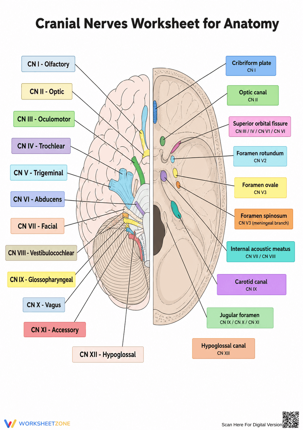
This high school anatomy worksheet provides a comprehensive visual guide to the twelve cranial nerves and their corresponding skull foramina. By studying this detailed diagram, students will map the pathways of the nervous system, connecting specific nerves to their anatomical exit points to better understand human biology.
At a Glance
- Grade: 9-12 · Subject: Biology
- Standard:
HS-LS1-2— Illustrate interacting systems providing specific functions in organisms- Skill Focus: Identifying cranial nerves and foramina
- Format: 1 page · 22 identification points · No answer key needed · PDF
- Best For: Visual reference and study guide
- Time: 15–20 minutes
What's Inside
Inside this single-page resource, educators will find a fully labeled anatomical diagram splitting the brain and skull base. The left side identifies cranial nerves I through XII using distinct color-coding. The right side pinpoints the exact skull foramina, such as the jugular foramen, that each nerve passes through. This dual-view layout functions perfectly as a standalone study guide.
Zero-Prep Workflow
This resource is designed for immediate classroom implementation.
- Print (1 minute): Print the PDF in color or grayscale.
- Distribute (1 minute): Hand out the diagram as students enter the lab.
- Review (3 minutes): Walk through the color-coding system, showing how nerves correspond to foramina.
With under two minutes of prep time, this diagram is an excellent addition to any biology sub plan.
Standards Alignment
Aligned to HS-LS1-2: Develop and use a model to illustrate the hierarchical organization of interacting systems that provide specific functions within multicellular organisms. Students visualize how the nervous and skeletal systems interact. Both standard codes can be copied directly into lesson plans, IEP goals, or district curriculum mapping tools.
How to Use It
This diagram serves as an excellent reference tool during direct instruction on the peripheral nervous system. Alternatively, it works perfectly as a self-study tool before a lab practical. For a quick formative assessment, have students cover the text boxes with sticky notes and attempt to recall the names from memory. Expected study time ranges from 15 to 20 minutes.
Who It's For
Designed for high school biology, AP biology, and introductory anatomy students. The clear visual scaffolds make it accessible for visual learners. It pairs perfectly with 3D skull models or interactive dissection labs, providing a reliable reference to support hands-on learning.
Mastering complex anatomical structures requires clear, accurate visual models that effectively reduce cognitive load for learners. This resource directly supports HS-LS1-2 by helping students illustrate interacting systems providing specific functions in organisms. According to Fisher & Frey (2014), providing students with high-quality, labeled visual scaffolds significantly improves their ability to retain complex domain-specific vocabulary and understand intricate spatial relationships in biological systems. By explicitly linking the twelve cranial nerves to their corresponding skull foramina in a single, color-coded diagram, this worksheet minimizes the split-attention effect often found in dense anatomy textbooks. Students can efficiently trace the pathways of the peripheral nervous system, reinforcing their understanding of how structural anatomy dictates physiological function. This targeted visual approach ensures that learners build a robust foundational schema, preparing them for advanced medical, nursing, or biological studies.