



















9th Grade Spinal Cord Worksheets
These 9th grade spinal cord worksheets resources give biology and anatomy teachers a set of printable activities covering spinal cord structure, neural pathway direction, reflex arc mechanics, and the functional consequences of spinal injuries — all in formats students can label, annotate, and complete as standalone practice. The set moves from macroscopic anatomy, including the meningeal layers and five vertebral regions, into cross-sectional structure, where students identify gray matter horns, white matter tracts, and the central canal. Reflex arc activities add a functional layer, so students work through the anatomy in motion rather than treating it as a static inventory of terms.
The Specific Skills Targeted in Each Worksheet
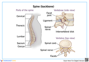
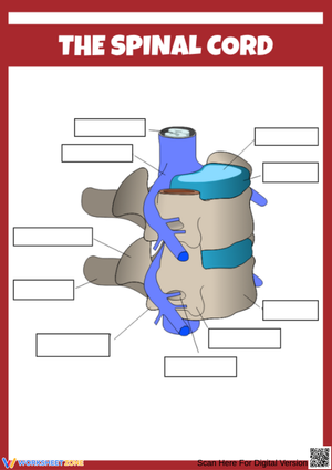
Each worksheet in the 9th grade spinal cord worksheets set moves deliberately from broad to detailed. Students start with full-body diagrams to locate the spinal cord relative to the brainstem and peripheral nervous system, then identify the five vertebral segments — cervical, thoracic, lumbar, sacral, and coccygeal — alongside the 31 spinal nerve pairs that exit at each level. That regional overview matters before students move into cross-sectional work; without a spatial sense of segmentation, they treat the cord as a single undifferentiated tube rather than a relay structure with distinct functions at different levels.
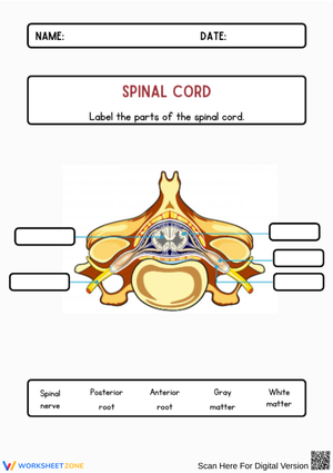
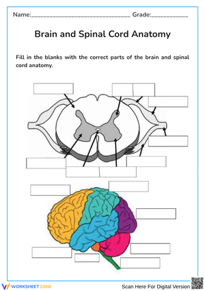
Cross-section labeling forms the anatomical core of the set. Students mark the three meningeal layers — dura mater, arachnoid mater, and pia mater — along with the subarachnoid space where cerebrospinal fluid circulates as a hydraulic buffer against trauma. Inside the cord, they identify the butterfly-shaped gray matter by labeling the dorsal horns (sensory input), ventral horns (motor output), gray commissure, and central canal. The white matter section asks students to distinguish ascending sensory tracts from descending motor tracts — a distinction that becomes clinically meaningful once students understand why injury at a specific vertebral level produces predictable, not random, loss of function.
Reflex arc activities complete the set. Students trace the full pathway — sensory receptor, sensory neuron, interneuron in the spinal cord, motor neuron, effector muscle — using flowcharts that require sequencing each step without routing the signal through the brain. A closing section distinguishes somatic reflexes involving skeletal muscles from autonomic reflexes governing smooth muscle, cardiac tissue, and glands.
Frequent Student Errors Worth Watching For and Correcting
The gray matter versus white matter distinction fails in a consistent way at this level. Most 9th graders arrive with the intuition that gray signals processing and white signals transmission — essentially the correct relationship — but they apply it spatially backward on cross-section diagrams, marking the outer white matter as the region containing neuronal cell bodies and placing the myelinated axons inward. The butterfly silhouette of gray matter in these diagrams gives students enough visual feedback to catch the mismatch themselves: if the cell-body label lands on the smooth outer ring rather than the butterfly outline, something is wrong.
Reflex arc tracing produces the most persistent error in the entire unit. Students understand that reflexes are fast and involuntary, but they still route the signal through the brain before the motor response fires — because every prior neural pathway they've learned has followed exactly that route. Ask a class to trace a hot-stove withdrawal reflex and a substantial portion will write: sensory neuron → spinal cord → brain → spinal cord → motor neuron → muscle. The flowchart format in these worksheets blocks that insertion structurally: the step after "interneuron in spinal cord" must connect directly to "motor neuron," with no brain-labeled node available. Students who make the error on an open-ended diagram stop making it on the flowchart, which gives a clean before-and-after comparison for formative assessment.
A third error shows up in how students represent directionality on cross-section diagrams. Students who correctly state that dorsal roots carry sensory signals will still draw arrows pointing away from the cord on the dorsal side, conflating "incoming signal" with "outgoing direction in the diagram." These worksheets use consistent arrow conventions — incoming on dorsal, outgoing on ventral — across multiple exercises, so students practice the directional logic in context rather than relying on a single memorized statement.
Fitting These Worksheets Into Your Teaching Week
The most effective sequence for the 9th grade spinal cord worksheets set puts the regional anatomy worksheet at the start of the nervous system unit — day one or two, before vocabulary density increases. Students need a spatial sense of where the spinal cord sits relative to the brainstem and the peripheral nerves before they encounter terms like arachnoid mater or gray commissure. Trying to build that spatial understanding retroactively, once students are already managing new vocabulary, adds unnecessary cognitive load at the wrong moment in the unit.
Cross-section labeling worksheets work well on days three through five, once students have seen the cross-section in direct instruction but haven't yet been assessed on it. A technique that consistently pays off: during the first labeling pass, have students assign warm colors — red and orange — to sensory structures (dorsal roots, dorsal horns) and cool colors — blue and purple — to motor structures (ventral roots, ventral horns). Students who color-code while completing the labeling recall directional flow more accurately on unit tests than students who work in a single color, because the distinction becomes spatial rather than purely verbal.
Reflex arc worksheets land well as the opening activity at the start of a class period — roughly the first eight minutes, before direct instruction begins. Give small groups different stimulus scenarios — stepping on broken glass, a patellar tendon tap, contact with a hot surface — and have them map the arc on a whiteboard before each student completes the flowchart individually. The group step surfaces the brain-routing misconception before whole-class instruction begins, so the lesson can address it directly using examples students just worked through themselves.
The injury-level worksheet fits near the unit's end. After students understand vertebral regions and their associated nerve pairs, asking them to predict the functional difference between an injury at C4 versus one at L2 gives immediate application to everything they've labeled across the unit. Students who initially found vertebral region naming arbitrary suddenly see why the cervical-versus-lumbar distinction matters in a medical context — and that realization tends to sharpen retention heading into the exam.
Adjusting the Set for a Range of Learners
The 9th grade spinal cord worksheets resources work across a range of preparation levels with targeted adjustments. For students who need vocabulary support, provide a labeled word bank alongside the cross-section diagrams — not a completed diagram, but a bank of terms that narrows the choice space without removing the labeling task. Students managing heavy working memory demands can concentrate on placing terms correctly rather than simultaneously recalling spelling and anatomical location.
For students who move through the standard labeling exercises quickly, the most effective extension is removing the word bank and asking them to add a brief functional annotation beside each label — not just "dorsal horn" but "dorsal horn: receives sensory input from peripheral receptors." That added step requires students to translate labeled anatomy into function, which surfaces gaps in understanding that the labeling task alone would miss.
Reflex arc worksheets offer a natural extension layer without requiring entirely separate materials. Students ready for more complexity can annotate each flowchart step with the type of neuron involved, the direction of signal travel relative to the central versus peripheral nervous system, or the neurotransmitter released at each synapse. These additions fit in the margins of the existing worksheet format, so the same resource serves multiple readiness levels within a single class period.
Frequently Asked Questions
What is the difference between the spinal cord and the vertebral column?
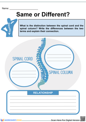
The spinal cord is the neural tissue itself — a cylindrical bundle of nerve fibers running from the base of the brainstem to approximately the first lumbar vertebra, and a core component of the central nervous system. The vertebral column is the bony structure of 33 stacked vertebrae that encases and protects the cord from mechanical injury. Students frequently use both terms to mean the same structure, which creates consistent confusion when injury discussions specify a vertebral level and students cannot distinguish the bone from the neural tissue inside it.
How does a reflex arc bypass the brain and still produce a coordinated response?
The spinal cord handles the entire reflex loop locally. A sensory neuron delivers the incoming signal to the spinal cord, where interneurons in the gray matter relay a motor command without waiting for the signal to travel to the brain and return. The brain receives notification of the reflex after it has already occurred — awareness follows action, not the other way around. This arrangement eliminates the roughly 20 to 40 milliseconds a full cortical loop would require, which is enough time to meaningfully reduce tissue damage from a harmful stimulus.
Why does the gray matter form a butterfly shape in cross-section?
The butterfly outline reflects the spatial organization of neuronal cell bodies and unmyelinated fibers within the cord. The upper wings are the dorsal horns, which receive incoming sensory signals from peripheral receptors. The lower wings are the ventral horns, which house the motor neuron cell bodies that send signals out to skeletal muscles. The gray commissure connects the two sides across the midline, and the central canal runs through its center. That arrangement — two pairs of horns joined by a commissure — produces the H or butterfly silhouette visible in any cross-section diagram.
What does cerebrospinal fluid actually do for the spinal cord?
Cerebrospinal fluid circulates through the subarachnoid space between the arachnoid mater and the pia mater and performs three distinct functions. It acts as a hydraulic shock absorber, reducing the mechanical impact of sudden movement on the neural tissue. It maintains the stable ionic environment the spinal cord requires for reliable electrical signaling. And it carries metabolic waste away from neural tissue — a drainage role the cord cannot perform through standard vascular channels. Students typically reduce this to "cushioning," which names only one of the three functions and misses the chemical stability and waste-clearance roles entirely.
Clear All