9th Grade Skull Anatomy Worksheets Printable
These 9th grade skull anatomy worksheets printable cover both cranial and facial bones through labeled reference diagrams and blank identification diagrams drawn from anterior, lateral, and superior views. Freshmen hit skull anatomy as one of the first genuinely vocabulary-dense units in high school biology — 22 bones, four named sutures, and a web of functional relationships that have to stick before the unit exam arrives. Diagram practice is where that retention actually happens, and this set gives teachers enough format variety to drive it across multiple days of instruction.
The Specific Bones and Structures Each Worksheet Covers
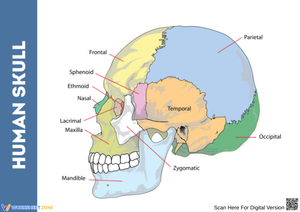
The eight cranial bones anchor every other skull discussion, so most worksheets in the set begin there. Students mark and name the frontal bone, the paired parietal and temporal bones, the occipital bone at the posterior base, and the sphenoid and ethmoid bones visible only in superior or internal-view diagrams. Functional notes appear alongside the diagrams — the foramen magnum on the occipital, the "keystone" role of the sphenoid on the cranial floor — because 9th graders retain anatomical names far better when the structure means something beyond a label to memorize.
Facial bone coverage spans all 14 structures:
- Mandible and maxilla — the jaw pair most critical for mechanical digestion, with the mandible being the skull's only freely movable bone
- Zygomatic bones — the cheekbones that also form the lateral walls of the eye orbits
- Nasal and lacrimal bones — small rectangular structures students routinely overlook on anterior-view diagrams
- Palatine bones and vomer — posterior hard palate and nasal septum structures that only read clearly in inferior or internal views
- Inferior nasal conchae — often the last bones freshmen master, because they're not visible from the surface
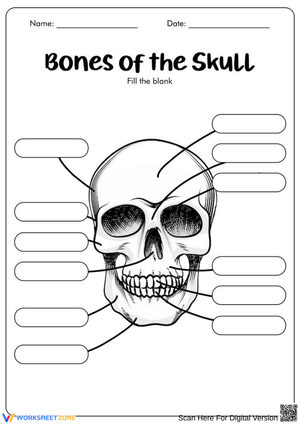
Students treat facial bones as secondary material, but they carry a disproportionate share of terminology points on lab practicals. Worksheets that pair an anterior cranial diagram with a facing anterior facial diagram help students see the two groups as an integrated system rather than two separate lists to memorize in isolation.
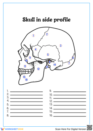
Sutures appear on lateral-view worksheets where the coronal, sagittal, lambdoid, and squamous lines are clearly drawn. One worksheet pairs the suture diagram with a brief structural note about how fontanelles allow cranial expansion during infancy — a direct structure-function connection that gives the lesson a developmental dimension and opens a genuine classroom conversation about how bone anatomy changes across a human lifespan.
Student Errors Worth Catching Before the Unit Test
The most reliable error in this unit is partial knowledge of the frontal bone. Students correctly name it across the forehead but miss that it also forms the superior margin of the eye orbits. On anterior-view diagrams, that gap surfaces as mislabeled orbital borders — a student writes "zygomatic" where the frontal actually extends, or leaves the superior orbit blank entirely. It doesn't surface until the first graded diagram check if teachers aren't actively watching for it on low-stakes practice work.
Suture confusion runs a close second, specifically between the lambdoid and sagittal. Students memorize that the sagittal runs along the midline, but on a lateral view where the midline isn't the orientation anchor, they transpose the two. Running one worksheet strictly as a bell-ringer focused on just the posterior cranium — before it gets folded into a full-skull review — isolates that confusion before it compounds.
A third pattern appears with the sphenoid and ethmoid. Because neither is visible in a standard anterior or lateral view, students who perform well on surface-bone diagrams go blank when shown a cranial floor diagram. The superior-view and internal base-view worksheets in this set address that gap directly; they shouldn't be treated as optional extension material.
Lesson-Planning Strategies for Getting the Most From These Worksheets
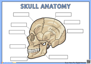
The most effective sequence is to open with a 3D model or a projected anatomical image, then hand students a labeled reference diagram the same day while the spatial memory is still fresh. The 9th grade skull anatomy worksheets printable that feature blank identification diagrams work best on days two and three of the unit, after students have already mapped the bones once. Spacing that blank-diagram practice over two or three separate sessions — rather than concentrating it all in a single review block — produces noticeably stronger recall by exam day. That's spaced retrieval doing its job, and these worksheets are built for it.
Bell-ringer use is practical and efficient. A blank lateral-view diagram takes most students around seven minutes to complete at the start of class, which fits the window between morning announcements and the main lesson without eating into instructional time. Exit tickets using a simplified three-bone or two-suture diagram give teachers a quick read on who is keeping pace and who needs a targeted reteach before the week closes.
For teachers who use interactive notebooks, the diagrams fold and glue into composition notebooks without trimming. Color-coding cranial bones in one hue family and facial bones in another — blues and greens for cranial, warm reds and oranges for facial — reduces sorting errors under test pressure and gives students a visual anchor when recalling structure location. Students who color the diagrams actively during a review session consistently outperform students who simply re-read labeled diagrams, because the coloring task forces them to trace each bone's boundary rather than skim past familiar labels.
Standard Alignment
These worksheets support NGSS HS-LS1: From Molecules to Organisms — Structures and Processes, specifically the expectation that students construct explanations for how the body's structural organization carries out essential life functions. Skull anatomy is an unusually direct fit for this standard because the structure-function relationship is visible and concrete: the thickness of the occipital bone protects the cerebellum; the lightness of the ethmoid supports its role in the nasal passages; the mandible's mobility enables mechanical digestion. Students are not inferring function from abstract molecular processes — they are reading it off a diagram.
HS-LS1 sits at the opening of the high school life science sequence, which is why skull anatomy so often anchors the freshman biology year. It establishes the habit of thinking in structure-function pairs before students move into cell biology, genetics, or organ-system units where those relationships are far less immediately visible. Diagram identification work — the primary task in this set — is also a direct preparation for the evidence-based argumentation practices NGSS asks students to develop throughout the high school band.
Adjusting the Set for a Range of Learners
Word-bank versions of each blank diagram give students who need extra support a manageable entry point. They are still doing identification and spatial matching rather than passive reading, but the retrieval demand is lower than cold recall from memory. Removing the word bank for students who are ready is the simplest tier adjustment available, and it takes no preparation beyond printing a different version of the same worksheet.
For students who move quickly through surface-bone identification, the internal base-view diagram presents a genuine challenge. The sphenoid's butterfly wings and the ethmoid's cribriform plate are disorienting in this perspective even for students who have anterior-view bones locked down. Pairing that worksheet with a short comparison task — "label the sphenoid here, then describe one thing visible in this view that the lateral view hides" — shifts the work from memorization to structural analysis.
Students with fine motor limitations sometimes struggle with the small bone outlines on detailed diagrams. Printing at 125 percent and using a simplified diagram that shows only the eight cranial bones keeps the cognitive work on anatomy rather than on handwriting mechanics. That adjustment also works as a starting point for students entering the unit with limited prior exposure to anatomical terminology.
Frequently Asked Questions
Which skull bones are most commonly assessed in 9th-grade biology?
Most freshman biology courses assess the frontal, parietal, temporal, occipital, sphenoid, maxilla, mandible, and zygomatic bones, along with the four major sutures. In more thorough anatomy units, the ethmoid, lacrimal, nasal, palatine, vomer, and inferior nasal conchae also appear. A teacher running a two-week skull unit typically introduces all 22 bones but holds students accountable for the eight cranial bones and six primary facial bones on identification assessments.
Do the diagrams include anterior, lateral, and superior perspectives?
Yes. All three major views appear across the set, because no single perspective shows every structure clearly. The anterior view is strongest for facial bone identification. The lateral view is where suture lines and the temporal bones read most clearly. The superior and internal base views are where the sphenoid and ethmoid finally make visual sense. Teachers who use 9th grade skull anatomy worksheets printable from only one perspective often find that students cannot transfer their knowledge when the orientation shifts on a test — which is precisely why having all three is necessary rather than supplemental.
Can a blank skull diagram work as a formative assessment, or is it only useful as practice?
It works well in both roles. A blank diagram used as an exit ticket at the end of a class period is a low-stakes formative check — teachers scan for consistent blank spots or recurring errors without building a full grading event around it. The same diagram used at the end of a unit, with all bones expected from memory, serves as a summative measure. The difference is entirely about timing and whether the score counts. The worksheet format is neutral enough to serve either purpose without any modification.
How does skull anatomy connect to later topics in the high school biology course?
The structure-function reasoning students build with the skull carries directly into the nervous system unit, where the cranial bones' relationship to specific brain regions becomes explicit. Students who understood why the occipital bone is thick have an easier time when the course covers cerebellar function. The mandible and maxilla reappear in nutrition and digestion units. An accurate spatial model of the skull built early in the year gives later units a concrete anatomical anchor rather than an abstract list of terms to re-introduce. These 9th grade skull anatomy worksheets printable do that foundational work up front — the more thoroughly students map the skull in the first unit, the less reteaching is needed when those bones resurface in different biological contexts later in the course.
Clear All