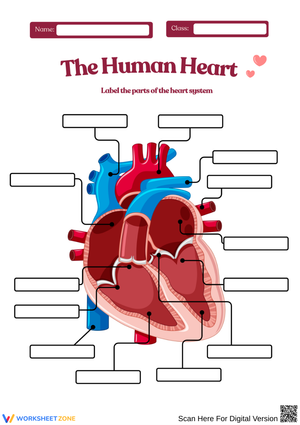
Comprehensive Heart Anatomy Worksheets for 9th Grade Biology
These heart anatomy worksheets for 9th grade cover the four chambers, all four valves, the major connecting vessels, and the full two-circuit blood flow sequence — in the exact progression teachers need for a unit that builds from identification toward mechanism. Each worksheet addresses a distinct layer of the topic, so teachers can assign them in sequence or pull individual worksheets to fill specific gaps before a lab or assessment.
What Each Worksheet Covers
The content moves through three levels that 9th grade biology requires: structural identification, spatial relationships within the heart, and function-based explanation. Students who work through the set in order are not just memorizing names — they are building a working model of how the heart actually moves blood.
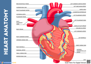
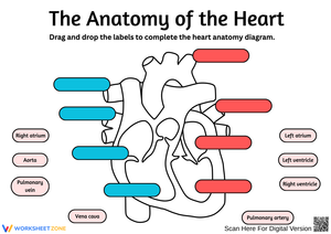
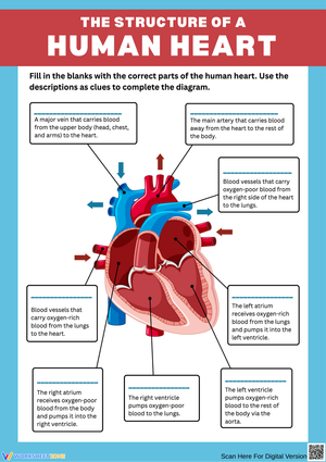
- The four chambers — right atrium, left atrium, right ventricle, left ventricle — with questions about both location and functional role (receiving versus pumping)
- The four valves — tricuspid, pulmonary, mitral, and aortic — grouped by type (atrioventricular versus semilunar) rather than listed in isolation, so students understand the structural logic
- Major vessels — superior and inferior vena cava, pulmonary artery, pulmonary veins, and the aorta — with attention to oxygenation status and direction of flow at each point
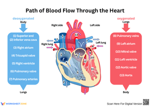
- Pulmonary and systemic circuit tracing — students follow a red blood cell through the full loop, labeling each stop and indicating whether the blood is oxygenated or deoxygenated
- Structure-function relationships — including the wall thickness difference between the left and right ventricles, explained in terms of pumping distance and pressure
Mistakes Students Make That Catch Teachers Off Guard
The most reliable error in this unit involves the pulmonary artery. Students who correctly memorize "arteries carry oxygenated blood" will shade the pulmonary artery red on every diagram — and feel confident doing it. The exception does not feel like an exception to them; it feels like confirmation of the rule they just learned. Simply restating the rule does not fix it. Students need to practice tracing deoxygenated blood from the right ventricle outward, step by step, before the distinction sticks. These heart anatomy worksheets for 9th grade include pathway-tracing tasks specifically because that active sequencing is what breaks the false generalization.
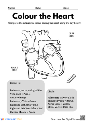
Two other errors appear consistently in student work. The first is orientation: on a standard frontal diagram, the anatomical right side of the heart appears on the viewer's left — the same mirror-flip that trips students up in every anatomical illustration. Students who have not been warned will label the right atrium on the right side of the page, and they will do it with complete confidence. Worth flagging explicitly before the first diagram task. The second error is functional: students can recite "atria receive, ventricles pump" but cannot apply it under pressure. Ask a student why the left ventricle wall is thicker and many will say it is "more important" rather than connecting wall thickness to pumping distance and the pressure demands of systemic circulation.
Building These Worksheets Into Your Biology Unit
The strongest placement for these worksheets is as pre-lab preparation. Before a sheep heart dissection — or a virtual simulation — students who can already identify the major structures on paper spend lab time observing and asking questions instead of flipping back to their textbook for basic names. Assigning the identification worksheets one or two days before the dissection means the lab itself becomes a moment for verification and comparison, not first exposure.
For daily lesson use, the color-coding task earns its time. Give students two colored pencils and have them trace deoxygenated blood in blue and oxygenated blood in red through a full heart diagram. This single task generates more discussion about the pulmonary circuit exception than most direct instruction does — the moment students reach the pulmonary artery and have to decide between blue and red is exactly when the concept lands. Heart anatomy worksheets for 9th grade that include this tracing task work especially well as a formative check: the coloring itself reveals where a student's mental model breaks down, often more clearly than a written quiz does.
For the broader unit arc, the most productive sequence is direct instruction on each substructure, then immediate worksheet practice, then a review block before any quiz. A Friday period spent on an unlabeled diagram — students labeling from memory before checking their work — builds retention far more reliably than re-reading textbook notes. Active retrieval is doing the work there, not the worksheet itself.
Standard Alignment
NGSS LS1.A: Structure and Function is the primary standard these worksheets address. At the 9th grade level, LS1.A moves beyond the general body systems overview that students encountered in middle school and into mechanism: students must explain why a structure is built the way it is, not simply name it. The left ventricle is not just "thicker" — students at this level are expected to connect that thickness to pumping distance, systemic resistance, and the pressure demands of delivering blood from the heart to the toes. Each worksheet keeps that structure-function expectation active, resisting the pull toward pure identification that makes heart anatomy feel like a vocabulary list rather than a biology concept.
Adjusting the Set for a Range of Learners
Students who are still building science vocabulary benefit from keeping a fully labeled reference diagram nearby while working — not as an answer key, but as a visual anchor. Limiting the initial task to the four chambers before introducing valves and vessels keeps cognitive load manageable and reduces the number of students who shut down midway through a complex diagram. For ELL students, a word bank with phonetic support on terms like atrioventricular and semilunar lowers the language barrier without reducing the content expectation.
Students who are ready to go further can move from identification into analysis: given the left ventricle's thicker wall, what would happen to systemic blood pressure if that muscle weakened? Which valve fails in mitral valve prolapse, and what does that mean for blood flow direction? These questions require the same anatomical knowledge as the base task but apply it to a clinical scenario — exactly where heart anatomy worksheets for 9th grade can stretch into honors-level thinking without requiring a separate document. The base worksheets carry the whole class; the extension questions add depth for students who finish early or are working in an advanced track.
Frequently Asked Questions
Why does the left ventricle have a thicker wall than the right?
The left ventricle pumps oxygenated blood out through the aorta into systemic circulation — every organ in the body, from the brain down to the feet. That distance, combined with the resistance of the full vascular network, demands sustained high pressure. The right ventricle only moves blood to the nearby lungs, which operate as a low-pressure circuit. The muscle mass on each side directly reflects the workload each side actually does, which is the structure-function relationship 9th grade biology asks students to explain, not just observe.
What is the key distinction between pulmonary and systemic circulation?
Pulmonary circulation is the short loop: right ventricle to lungs to left atrium. Its job is gas exchange — offloading carbon dioxide, picking up oxygen. Systemic circulation is the long loop: left ventricle out through the aorta to every tissue in the body, then back through the vena cava to the right atrium. One circuit oxygenates the blood; the other delivers it. Students who can state this clearly and trace it on a diagram without prompting have met the core expectation for this unit.
How do these worksheets work for both formative and summative assessment?
For formative use, assign a worksheet mid-lesson and circulate to check labeling accuracy before errors settle in. The blood flow tracing tasks are especially useful here — a glance at a student's color-coded diagram shows exactly where their understanding stops. For summative use, remove all labels from a diagram and ask students to identify structures, indicate oxygenation status at each point, and explain the function of one specific valve in a sentence or two. That combination — identification plus functional explanation — covers the full range of what a unit test would assess and gives teachers a clear picture of what each student actually knows.
Clear All