These muscles of the head and neck worksheets for 10th grade give biology teachers a focused set of resources for one of the anatomically densest regions students encounter in a body systems unit. The head and neck pack more named muscles into less surface area than almost any other region, and that density — layered over unfamiliar Latin-root terminology — is exactly what makes deliberate practice with labeled diagrams and functional groupings worth the class time.
Structures and Functions Covered Across the Set
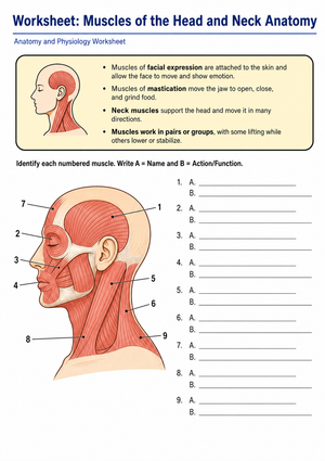
Each worksheet targets a specific functional group, which keeps cognitive load manageable at a stage where students are memorizing anatomical vocabulary for the first time. The three main groupings are muscles of facial expression, muscles of mastication, and the major muscles that move and stabilize the neck.
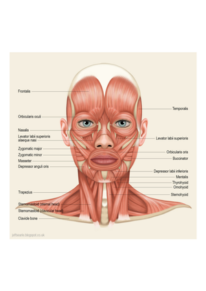
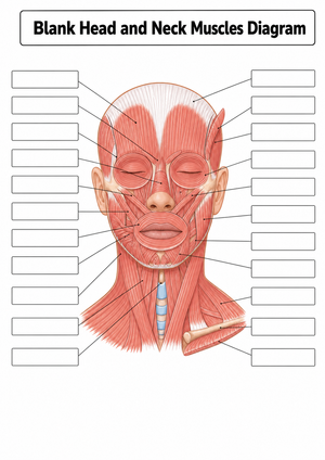
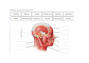
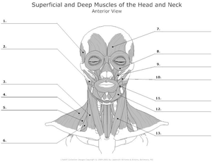
Facial expression muscles stand apart from most skeletal muscles because many insert directly into the dermis rather than attaching bone to bone. The frontalis, orbicularis oculi, orbicularis oris, and zygomaticus major appear across multiple worksheets, with diagrams showing their unusual skin-to-muscle attachment patterns. Students label each muscle and write a brief functional description, which pushes them past simple identification toward understanding why that particular structure produces that particular movement.
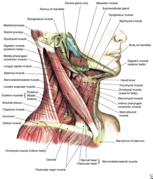
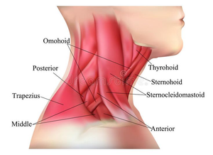
The mastication group is smaller but biomechanically rich. The masseter and temporalis appear on every worksheet in this group, with origin-insertion labeling tasks that ask students to predict the direction of pull before confirming the answer. The neck worksheets concentrate on the sternocleidomastoid and upper trapezius — both visible in lateral-view diagrams and palpable on a student's own body, which makes the anatomy suddenly concrete in a way that diagrams alone cannot.
What Sets This Region Apart Anatomically
The skin-insertion characteristic of facial muscles is worth dwelling on in class because it directly challenges the bone-to-bone lever model students just spent time building. When a student asks why the orbicularis oris can purse lips rather than pivot a joint, the answer is that the muscle fiber is pulling skin tissue — no lever, no fulcrum in the traditional sense. That distinction also opens a natural door to clinical conversations: Bell's palsy, temporomandibular joint dysfunction, and tension headaches caused by chronic frontalis contraction are conditions many 10th graders have encountered or heard about, and the clinical hook reliably increases engagement during what might otherwise feel like a memorization exercise.
How to Build These Worksheets Into Your Lesson Plans
The most reliable use pattern is pre-lab preparation. Before students handle physical anatomical models or run through a virtual dissection, one worksheet from this set orients them to the terminology and spatial layout. Students who arrive at the model having already labeled a diagram find structures faster and ask more specific questions — the worksheet does the orientation work so model time stays focused on three-dimensional relationships rather than basic name recall.
Color-coding tasks built into several worksheets work well during a structured 20-minute work block. Assigning one color per functional group — mastication muscles in one color, facial expression in another, neck movers in a third — produces a visual map students actually reference during later review. The finished diagram also functions well as an exit-ticket format: ask students to point to any muscle and state one action before they leave. These muscles of the head and neck worksheets for 10th grade pull double duty as Monday warm-ups at the start of a body systems review week, giving students a low-stakes retrieval task before a unit assessment without requiring new instruction time.
Frequent Student Errors Worth Watching For and Correcting
The most consistent error in student work is misidentifying the temporalis. Students frequently place it lower on the skull — sometimes labeling it directly on the zygomatic arch — because the word "temporal" suggests a narrow strip near the temple rather than a broad fan spreading across the parietal and temporal bones. Worksheets with a large lateral-view diagram surface this error immediately: if a student's label line points to the arch, they have the masseter location correct and the temporalis location wrong, which is a fast correction in the moment.
A second persistent error involves the sternocleidomastoid's action. Students who correctly memorize that it "rotates the head" almost universally write that it rotates the head toward the same side as the contracting muscle, when the action is actually rotation to the opposite side. Having students trace the muscle's diagonal path on the worksheet diagram — from the sternum and clavicle up to the mastoid process behind the ear — and then predict the direction of pull before committing to an answer catches this misconception before it gets embedded through repetition.
A third error is subtler: students confuse origin and insertion on facial muscles because the standard "fixed point" rule breaks down when one attachment is skin rather than bone. Worksheet tasks that ask students to mark the origin with a dot and the insertion with an arrow force them to reason through the anatomy rather than copy a label from memory.
Standard Alignment
These worksheets support NGSS HS-LS1-2, which asks students to develop and use a model to illustrate the hierarchical organization of interacting systems within multicellular organisms. When students label how the masseter connects the zygomatic arch to the mandible and then describe the nervous signal initiating that contraction, they are modeling exactly the muscular-skeletal-nervous interaction the standard requires. The labeling tasks serve as the modeling activity; the functional description questions address the interacting-systems component. Teachers working through a unit progression that moves from cell biology through tissues into organ systems will find this set fits cleanly at the end of the tissue unit and the opening of the organ systems sequence — a natural conceptual bridge between structure and integrated function.
Differentiating the Set Across Ability Levels
Students working below grade level do better when the worksheet provides a word bank and limits the scope to four or five muscles per diagram rather than twelve. Pulling a focused subset — the masseter, temporalis, orbicularis oris, sternocleidomastoid, and frontalis — and pairing it with a completed reference diagram gives those students a manageable entry point without stripping out the core anatomical vocabulary they still need to acquire.
Students ready for greater challenge can complete the origin-insertion labeling tasks without the word bank and then write a brief mechanical explanation for each muscle: what structures does it connect, what direction does it pull, and what movement results? A second extension is the clinical connection question included in several worksheets, which asks students to predict how damage to a specific nerve or muscle would alter a patient's facial movement. That reasoning task moves comfortably into AP Biology or pre-health elective territory while staying grounded in the same diagram every other student in the class worked from.
Frequently Asked Questions
Which muscles should 10th graders be able to identify and describe from memory?
The core set for a standard high school anatomy unit includes the masseter, temporalis, frontalis, orbicularis oculi, orbicularis oris, zygomaticus major, buccinator, sternocleidomastoid, and upper trapezius. That covers the major functional groups and represents the structures most commonly assessed on introductory high school anatomy tests. Students who can name each muscle, state its primary action, and place it in its functional group are well-positioned for a unit exam.
Can these worksheets replace anatomical models?
These muscles of the head and neck worksheets for 10th grade prepare students for model work — they don't replace it. Two-dimensional diagrams establish terminology and spatial relationships in a low-stakes setting before students interact with the model. The way the temporalis fans across the lateral skull only becomes fully clear when students rotate a physical or digital model and see the structure from multiple angles. The worksheet handles the vocabulary; the model adds the dimension the worksheet cannot.
How much class time do these worksheets typically require?
A labeling worksheet with a word bank runs about 12 to 15 minutes for most 10th graders working independently. An origin-insertion task with functional description questions takes 20 to 25 minutes. Coloring worksheets covering the full lateral diagram of the head can stretch to 30 minutes when students are being thorough — those work better as structured work time than as quick warm-ups.
Do students need prior anatomy instruction before starting this set?
These worksheets assume students can read a lateral-view anatomical diagram and understand basic directional terms — superior, inferior, anterior, posterior. Students who arrive without that background need a brief orientation to anatomical position before the worksheet makes full sense. A five-minute introduction to standard anatomical views removes that barrier cleanly and takes nothing away from students who already have the foundation.