These fish anatomy worksheets pdf give middle school science teachers clean, print-ready diagrams with label lines that land unambiguously on the right structures — not approximate, not crowded — and a difficulty sequence that tracks the natural arc of a vertebrates unit. The set moves from orienting students on the fish's body surface to identifying internal organs within functional systems, which matches how most life science textbooks progress and how dissection labs unfold in real classroom time. Teachers who plan perch dissections find the worksheets cut prep time and significantly reduce the disorientation students experience when they first encounter an actual specimen.
The Specific Structures in Each Worksheet
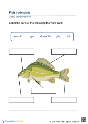
Each worksheet targets either external features or internal organ systems without conflating the two, because mixing them before students have orientation on the body surface reliably generates confusion. The external worksheets cover:
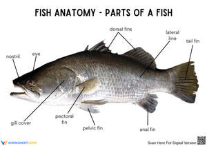
- Fins: dorsal, caudal, anal, pectoral, and pelvic — with enough diagram detail that students can distinguish unpaired midline fins from paired fins by position, not just by memorized label
- Operculum: the bony gill cover that students almost universally call "the gills" on first encounter, making it worth its own pointed diagram emphasis
- Lateral line: the faint sensory stripe running from head to caudal fin, present on virtually every state assessment covering vertebrate adaptations
- Scales and nares: treated together as surface structures, with brief functional notes printed alongside each diagram so students connect form to function from the start
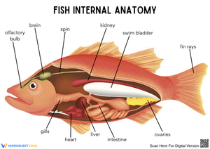
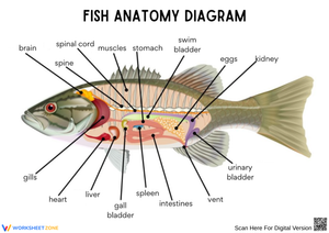
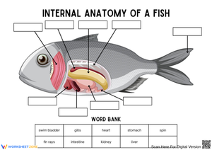
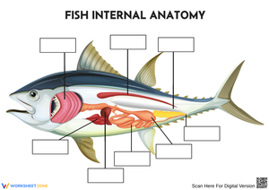
The internal worksheets move organ by organ through the major systems. Students label the two-chambered heart, trace the gill arches responsible for gas exchange, identify the swim bladder and its function in buoyancy regulation, and locate the digestive structures — stomach, liver, and pyloric caeca — before encountering the gonads. The sequence is deliberate: respiratory structures first, then circulatory, then digestive, because that order follows the conceptual thread from "how does it breathe" to "how does it fuel itself," building each system on the one before it.
The Grade-Level Logic Behind This Topic
Fish anatomy typically enters the curriculum in fourth grade as part of a unit on how structures support organism survival, then reappears with greater physiological depth in sixth through eighth grade when students study body systems, cell specialization, and comparative vertebrate anatomy. Bony fish appear at both levels for the same reason: the organ count is manageable, the structures are visually distinct from one another on a diagram, and the functions connect in ways students can reason through without advanced chemistry background. A fish anatomy worksheets pdf set designed for fourth grade leans on matching and coloring tasks; the same topic at seventh grade demands written function explanations and system-level comparisons. These worksheets support both contexts — the diagrams are clean enough for a ten-year-old to read and detailed enough to challenge a thirteen-year-old who already knows the label names.
Student Errors Worth Catching Before Students Reach the Lab
The most consistent error on external diagrams is anal fin mislabeling. Students who correctly place the dorsal fin along the top will rotate their spatial reasoning and write "second dorsal fin" for the unpaired fin below the body, because both are midline fins and the diagram's orientation doesn't always make the ventral surface obvious. Stating explicitly that the anal fin sits on the posterior-ventral surface — before students begin the worksheet — prevents a round of corrections that eats into class time.
On internal diagrams, the swim bladder and the stomach generate predictable confusion because both appear as elongated sacs in the mid-body region of a two-dimensional cross-section. Students who answer correctly are using dorsal-ventral positioning; students who flip them haven't registered that spatial cue. Pointing out the swim bladder's position above the digestive tract before students start is more effective than untangling the error afterward, when the misconception has already been written down and reinforced.
A third pattern appears specifically when students move from external to internal work: they expect to find the gills as visible red structures and become confused when the internal diagram shows gill arches instead. Students have learned "gills" as a single noun, so the idea that gills are a multi-part structure — filaments attached to arches, covered externally by the operculum — needs explicit framing before the internal worksheet begins.
Fitting These Worksheets Into Your Weekly Plans
The most effective placement for this set is a two-day pre-lab before a perch dissection. On day one, students complete the external diagram as a whole-class activity, with the teacher projecting a specimen photograph alongside the worksheet so students connect the line art to a real fish. On day two, the internal worksheet runs as partner work, with students defending their label placements to each other before answers are confirmed. When the dissection runs on day three, students arrive with anatomical vocabulary and spatial expectations already established — they're not discovering the swim bladder exists for the first time while holding a scalpel.
Outside of dissection prep, the external worksheet works well as a Monday warm-up during a vertebrates unit: a five-minute retrieval task that costs almost no instructional time but reinforces fin vocabulary from the previous week's reading. A fish anatomy worksheets pdf also drops cleanly into a stations rotation — one station for external labeling, a second for a short reading passage on countercurrent exchange in gills, a third for a swim bladder buoyancy matching task. The rotation format prevents the passive attention drift that sets in when anatomy instruction runs entirely through lecture.
Adjusting the Work Across Ability Levels
For students who struggle with spatial orientation on diagrams, the most practical adjustment is providing a word bank and reducing the label count on the first external worksheet. Asking those students to accurately place eight structures before introducing the remaining four gives them a foothold without stripping the diagram entirely — fish still have a lateral line even if it's held back for the second pass. Accuracy on a smaller set matters more than incomplete labels across all structures.
Students who move through labeling quickly benefit from an extension that asks them to color-code by system: respiratory structures in one color, digestive in another, circulatory in a third. The same diagram that served as a labeling task now requires a conceptual layer — students can't color the operculum as a respiratory structure without reasoning about its role in moving water over the gills. A further challenge asks them to draw arrows indicating the direction of water flow through the operculum and gas exchange across the gill filaments, converting a labeling exercise into a process diagram that demands system-level thinking.
Standard Alignment
The external anatomy worksheets address NGSS 4-LS1-1, which requires students to construct arguments that plants and animals have internal and external structures functioning to support survival, growth, behavior, and reproduction. At fourth grade, labeling the operculum, lateral line, and fin types directly builds the evidence students need for that argument — they aren't accumulating vocabulary in isolation but linking each structure to a specific function. For middle school, the internal organ worksheets extend into NGSS MS-LS1-3, which focuses on how body organization enables chemical energy conversion. Tracing the digestive organs — stomach, pyloric caeca, intestine, liver — and connecting them to how the fish acquires and processes energy satisfies this standard's demand for system-level reasoning rather than isolated identification.
Frequently Asked Questions
Are these worksheets appropriate for fourth grade, or do they target middle school?
Both grade bands work, but with different worksheets from the set. The external anatomy diagrams — fin identification, operculum labeling, lateral line recognition — fit comfortably into a fourth-grade life science unit on organism structures. The internal organ worksheets, especially those requiring written explanations of swim bladder function and gill exchange mechanics, align more cleanly with grades 6–8. A fish anatomy worksheets pdf set is most useful when teachers assign external diagrams to younger students and reserve internal system work for middle school.
How do these worksheets connect to a perch dissection lab?
They function as pre-lab preparation. Students who complete the external and internal labeling activities before the dissection arrive knowing where to look and what to call what they find. Without that preparation, a meaningful portion of lab time disappears into basic orientation — students trying to figure out which fin is which while the specimen sits in front of them. The worksheets and the dissection teach the same content; the worksheets make the dissection more productive by handling identification before the lab period begins.
Can I use these worksheets without a real fish specimen?
Yes. Photographs and short video walkthroughs work well alongside the diagrams, particularly for external features. Projecting a close-up photograph of a perch's operculum beside the worksheet line drawing helps students connect diagram abstraction to what an actual fish looks like. For internal structures, a labeled dissection photograph or a brief video passage through the body cavity accomplishes the same purpose. Students who have seen the actual structures — even in photographs — label diagrams with noticeably more accuracy and confidence than students who have worked only from line art.