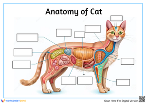
Cat Anatomy Worksheets PDF: Printable Labeling Activities for Biology Class
These cat anatomy worksheets pdf give biology teachers a print-ready collection of labeled diagrams covering every major body system — skeletal, muscular, digestive, respiratory, cardiovascular, and urogenital — that works equally as pre-dissection preparation, a cruelty-free alternative to a physical specimen, and a systematic review sequence before a unit exam. The diagrams reproduce cleanly in black and white, require no lab materials, and fit naturally into high school biology, anatomy and physiology electives, and introductory veterinary science courses.
Body Systems and Structures in the Set
The skeletal worksheet maps the feline's roughly 244 bones, including structures students rarely encounter in standard human anatomy units — the free-floating clavicle and the unusually long lumbar spine adapted for the cat's characteristic range of motion. That structural difference matters instructionally: students who understand why a cat's spine moves differently from a human's have internalized bone-function relationships, not just memorized label positions.
The remaining worksheets cover:
- Muscular system: Superficial and deep muscle layers from both dorsal and ventral views, with origins, insertions, and primary actions labeled on separate diagrams to reduce visual crowding.
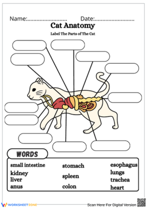
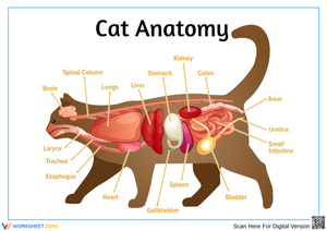
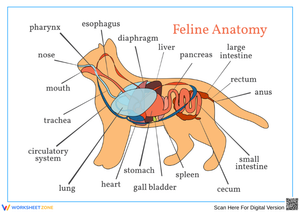
- Digestive system: Oral cavity through large intestine, with close-up insets of the stomach and ileocecal junction — structures students reliably confuse when viewing a full-body diagram.
- Respiratory system: Trachea, bronchi, left and right lung lobes, and diaphragm, with directional airflow indicators included.
- Cardiovascular system: Heart chambers, major vessels, and color-coded indicators distinguishing oxygenated from deoxygenated blood.
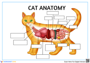
- Urogenital system: Kidney, ureter, bladder, and reproductive structures, with both male and female diagrams included.
Pre-Lab Labeling and the Logic Behind It
The cognitive case for diagramming before dissection is straightforward: when students have already named a structure, they recognize it in three dimensions instead of searching for it while the clock runs. Teachers consistently report that students who complete the corresponding worksheet the night before a dissection period get twice as far into the specimen as students who arrive unprepared — not because the prepared students are faster, but because they spend their lab time observing and analyzing rather than hunting for vocabulary in their notes.
Structured labeling also does something lecture alone cannot: it forces a commitment. A student who isn't sure whether the mass beneath the stomach is the pancreas or the spleen must decide on paper, and that decision — right or wrong — creates a memory trace that the lab experience either confirms or corrects. That confirmation-or-correction loop is more durable than passive note-taking, and it's the main reason pre-lab diagramming works even for students who initially label structures incorrectly.
For schools that restrict dissection, or for students who opt out, each cat anatomy worksheets pdf in the set pairs directly with high-resolution dissection photography and virtual tools. Distributing a worksheet alongside a dissection photograph and asking students to label from the image replicates the observation-analysis cycle without a physical specimen. Adding a short-answer column — "Explain the function of this structure in one sentence" — keeps the task analytical rather than purely reproductive.
Frequent Labeling Errors Worth Anticipating and Addressing
The most consistent pattern across student work is positional reasoning: students assign labels based on where a structure sits relative to others on the page rather than what that structure actually is. On cardiovascular diagrams, a reliable majority correctly identify the heart but then label the pulmonary veins as arteries because they arrive at the heart from the top — the same positional slot where arteries depart in diagrams studied earlier. A brief direct comparison of vessel function versus vessel position, held at the start of the cardiovascular worksheet period, cuts this error sharply.
On the muscular worksheet, students confuse the sternomastoid and the brachiocephalic — two muscles that sit adjacent in the ventral neck view. This is almost never a vocabulary failure; it is a spatial orientation problem. Students who mislabel these structures are typically reading the diagram from the wrong reference point. Drawing a midline on the board and labeling anterior, posterior, medial, and lateral before students begin reduces the error rate noticeably and takes about 90 seconds of class time.
A third pattern: students who correctly label the cecum in the close-up inset worksheet will often transpose it with the ileum on the full-body diagram, because the relative scale is different from the view they practiced with. Requiring students to trace the path of a food bolus — naming each organ in sequence while pointing to it on the diagram — surfaces this confusion before it appears on a quiz.
Building These Worksheets Into a Two-Week Anatomy Unit
One body system per class period is the right pace for most high school groups. Open with a 10-minute vocabulary preview, distribute the worksheet for guided completion, and circulate while asking function-level questions rather than correction questions — "What does that vessel carry?" rather than "Is that right?" — because function questions expose positional reasoning errors before they become fixed. Collect worksheets at the end of the period and scan them before the next class to identify which structures need re-teaching.
The sequencing matters as much as the individual worksheets. Running the skeletal worksheet on Monday, the muscular worksheet on Wednesday, and a combined review on Friday — rather than covering all three in a single block — spaces retrieval attempts enough that students retain label positions across the unit rather than cramming and forgetting. Spaced retrieval practice is the research-supported reason this pacing works, and it is easy to build into the week when each system has its own worksheet.
Each cat anatomy worksheets pdf in the set also works as a pre-lab assignment. A five-question identification quiz at the start of the lab period, drawn directly from the worksheet completed the night before, sets the expectation that preparation matters and measurably shortens the time students spend locating structures during dissection.
Standard Alignment
These worksheets connect most directly to NGSS HS-LS1-2, which asks students to use models to illustrate the hierarchical organization of interacting systems within multicellular organisms. Each labeled diagram is exactly that kind of model — students identify a system's structures and then answer questions requiring them to explain what those structures do and how the system connects to adjacent systems. The standard's emphasis on "interacting systems" is why pairing the cardiovascular and respiratory worksheets in the same week produces stronger conceptual outcomes than treating them as separate units: gas exchange only makes sense as a cross-system event, and students need to see that relationship made explicit.
The broader disciplinary core idea supporting this work is LS1.A (Structure and Function), which expects students to connect structure to function at the organ and system level. Cat anatomy is a productive context for LS1.A because its structural specializations — the flexible lumbar spine, the compact digestive system proportioned for a carnivore's diet, the kidney features tied to water conservation — give students concrete cases where structure visibly explains function rather than requiring abstract argument.
Tailoring the Worksheets for Different Learner Levels
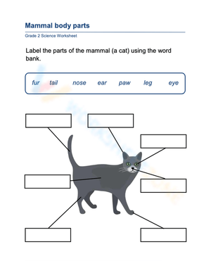
The built-in word banks make each worksheet accessible to students who are still acquiring anatomy vocabulary alongside content, but they limit the cognitive demand for students who already know the terms. The most efficient differentiation move is to print two versions of the same worksheet: one with the word bank intact for students working toward mastery, and one with the word bank removed for students ready to demonstrate recall. Both groups complete the same diagram; the difference in difficulty is significant without requiring separate lesson planning.
For students who struggle with anatomical orientation — dorsal/ventral, anterior/posterior — add a small reference card to their workspace before they begin. A diagram showing a cat in standard anatomical position with the four directional terms labeled removes that particular barrier without reducing the anatomical challenge itself. The goal is to direct their cognitive effort toward the structures rather than toward figuring out which side of the diagram is which.
Advanced students benefit from cross-system comparison tasks after completing two or more worksheets. Ask them to identify three locations where the arrangement of bones directly influences the attachment or action of a nearby muscle group, and to explain the mechanical relationship at each site in writing. This pushes well beyond labeling into the analytical reasoning that NGSS performance expectations actually require, and it connects naturally to the comparative anatomy conversations that make working with feline specimens — or their paper-based equivalents — worth doing in the first place.
Frequently Asked Questions
Are these worksheets appropriate for both dissection and non-dissection classrooms?
Yes. The diagrams work as pre-lab preparation in classrooms that include dissection, and they work as the primary anatomy resource in schools where dissection is restricted or where individual students opt out. Pairing each worksheet with a dissection photograph or a still from virtual dissection software provides the visual context a physical specimen would otherwise supply. The short-answer questions included with each system ensure that students who don't dissect still engage analytically with function, not just structure.
What grade level does this set target?
The vocabulary and diagram complexity target grades 9–12 — specifically high school biology, anatomy and physiology electives, and introductory veterinary science courses. The diagrams are detailed enough to challenge AP Biology students and clear enough to serve as first-exposure materials for ninth graders encountering body systems for the first time. Middle school science teachers covering basic organ systems have also drawn on individual worksheets from the set as extension materials for advanced students.
How does cat anatomy connect to human anatomy in the curriculum?
Feline and human anatomy share the same major body systems organized along the same developmental blueprint — which is why cats have served as anatomy models in secondary education for well over a century. The homologous structures between species give teachers a concrete comparison point: students who label both a feline femur and a human femur and then write a sentence explaining both the structural similarity and the functional difference are doing genuine comparative biology. A full set of cat anatomy worksheets pdf resources makes that comparison systematic and teachable rather than incidental, building the cross-species reasoning that students pursuing health sciences or veterinary careers will draw on throughout their training.
Do the worksheets include answer keys?
Each worksheet includes a fully labeled answer key. Having the key available makes it practical to use these worksheets as formative assessments — collect completed diagrams, compare against the key, note which structures were most often mislabeled, and use that pattern to plan re-teaching before the unit exam.
Clear All