Parts of a Leaf Worksheets PDF
These parts of a leaf worksheets pdf give 3rd-through-7th-grade science teachers print-ready diagrams and exercises spanning visible external structures and the microscopic cell layers that drive photosynthesis — the same anatomy that appears in NGSS performance tasks and end-of-unit assessments. Each worksheet targets a specific tier of leaf study, from naming the blade and petiole on a whole-leaf diagram to cross-sectioning the mesophyll and locating guard cells flanking individual stomata. Teachers who pair these with actual plant specimens consistently find that vocabulary holds longer when students can match a printed diagram against the real leaf in their hands.
Concepts Each Worksheet Targets
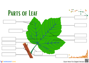
A well-structured parts of a leaf worksheets pdf covers two instructional phases that track how leaf anatomy deepens across grade bands. The external structure worksheets ask students to label seven to nine features on a whole-leaf diagram — blade (lamina), petiole, midrib, lateral veins, apex, margin, and base — then rewrite each term in a sentence connecting structure to function. That second step is where the real learning happens: a student who writes "the midrib carries water from the petiole outward into the smaller veins" understands something that a student who only placed a label does not.
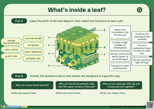
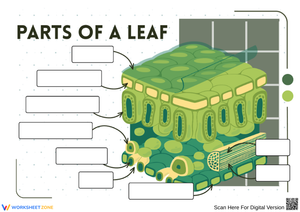
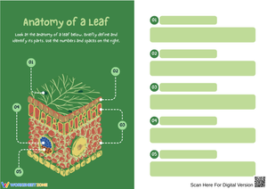
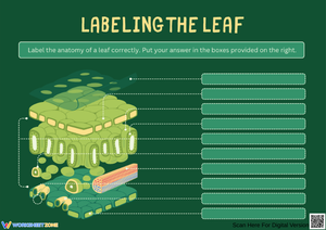
The internal cross-section worksheets shift to cellular anatomy and ask students to distinguish palisade mesophyll from spongy mesophyll, trace both the upper and lower epidermis, locate the cuticle, and mark individual stomata alongside their guard cells. A function-matching exercise accompanies each cross-section — students draw arrows from structures to biological roles:
- chloroplasts → light capture
- guard cells → regulating water loss
- xylem → upward water transport
- phloem → sugar distribution
Students who complete that exercise without a word bank have reached the level of understanding the standard actually requires.
Frequent Student Errors Worth Addressing Before the Lesson Starts
Several predictable errors appear in student work on leaf anatomy, and knowing them in advance lets teachers establish the right distinctions before confusion takes hold.
At the elementary level, the most persistent confusion is treating "petiole" and "stem" as interchangeable. Students who spent earlier grades hearing that the stem holds the plant up don't readily assign a separate name to the leaf stalk. A reliable correction: have students trace the water pathway from roots → stem → petiole → midrib → lateral veins on a whole-plant diagram, labeling each segment in sequence. Once the petiole is part of a continuous system rather than an isolated vocabulary term, the distinction holds through the assessment.
At the middle school level, the cuticle causes the most predictable trouble. Students routinely treat it as an internal tissue layer rather than a surface coating, and they collapse it into the epidermis directly beneath it. A short analogy — the cuticle is the wax on an apple's skin; the epidermis is the skin itself — takes about four minutes to establish and prevents most of that conflation before the worksheet goes out.
One more pattern to anticipate: when asked to mark stomata on a cross-section diagram, roughly half of a typical sixth-grade class places them on the upper epidermis. Correcting this during whole-group instruction — and explaining that concentrating stomata on the shaded lower surface reduces water loss during peak photosynthesis hours — turns a labeling correction into a functional insight. Students who understand why stomata are where they are remember the detail far longer than students who simply memorize the placement.
How to Work These Worksheets Into Your Lesson Plans
The external labeling worksheet works best on the day students first handle a physical leaf. Distribute the specimen before the worksheet — three to five minutes to observe and sketch independently — then hand out the diagram. This sequence lets students notice real structures first; the printed labels confirm what they saw and name what they couldn't identify. Running it the other way, worksheet before specimen, often turns observation into a scavenger hunt to match the diagram rather than genuine noticing.
Cross-section worksheets fit naturally alongside microscope lab days. When the lab isn't available, projecting the worksheet on a document camera and walking through each layer verbally creates a shared reference before students work independently. Either way, the function-matching exercise makes a strong exit ticket: six minutes to complete, and the results immediately show which students grasp the purpose behind each structure and which ones are still working at the level of label placement alone.
One classroom strategy that pays off quickly: have students color-code the vascular tissue before labeling — blue for xylem, yellow for phloem. This takes an extra three minutes but visually separates the transport structures from the photosynthetic tissue, reducing the confusion between veins and mesophyll that shows up in mid-unit checks. For Monday warm-up after a weekend gap, a blank diagram with no word bank — cold recall, no hints — is among the most efficient uses of the first eight minutes of class. The retrieval effort strengthens retention more than re-reading notes, and the results tell teachers exactly which structures need revisiting before the week's next lesson.
Standard Alignment
These worksheets address NGSS LS1.A: Structure and Function, which asks students to explain how an organism's internal and external structures support its survival, growth, behavior, and reproduction. At the elementary level, LS1.A performance expectations focus on connecting visible plant parts to observable functions — exactly what the external labeling and function-sentence exercises produce. At the middle school level, the same standard requires cellular-level explanation: students must account for how the arrangement of mesophyll layers, the distribution of stomata, and the presence of chloroplasts collectively support photosynthesis as a chemical process. A completed cross-section worksheet — where a student has labeled guard cells and written a sentence explaining their role in gas exchange — is direct, portfolio-ready evidence for a middle school LS1.A performance task. Any parts of a leaf worksheets pdf completed with the function-matching component also serves as a formative assessment artifact that documents student reasoning across the unit, which matters when building standards-evidence portfolios at the end of a grading period.
Adjusting the Set for a Range of Learners
For students working below grade level, keep the word bank visible throughout the task rather than removing it mid-exercise. Printing a brief glossary of the three most challenging terms — petiole, lamina, stomata — directly on the worksheet removes a vocabulary access barrier without lowering the conceptual demand. Students still place labels correctly and write function sentences; they simply aren't derailed by unfamiliar word forms before they can demonstrate their understanding of the anatomy.
Advanced students benefit from a stripped version of the cross-section worksheet: the structural outlines are present, but no labels or word bank appear. After labeling from memory, these students write a paragraph explaining how two specific structures interact — for example, how guard cells and the cuticle cooperate in a drought-adapted plant. That extension moves the task from identification into the mechanistic reasoning that appears later in AP Biology and introductory college science coursework.
For English language learners, projecting a bilingual leaf diagram while students work on the English-language worksheet supports vocabulary acquisition without reducing scientific rigor. The function-matching format is particularly accessible because it reduces the writing demand while still requiring students to demonstrate conceptual understanding — a meaningful distinction from simply copying a term onto a blank line.
Frequently Asked Questions
What grade levels are these worksheets designed for?
External structure worksheets are written for grades 3–5, where LS1.A first appears as a whole-plant performance expectation. Internal cross-section worksheets are designed for grades 5–7, when students begin reasoning about cellular structures they cannot observe directly — a shift that requires abstract inference that younger students find genuinely difficult, not merely unfamiliar. Each worksheet identifies its target grade band directly on the page.
Do these work alongside microscope labs, or as a standalone resource?
Both. On lab days, the cross-section worksheet gives students a reference structure to check their slide observations against — and they learn quickly that real tissue sections don't look as clean as printed diagrams, which is itself a valuable lesson about scientific representation. On non-lab days, the diagrams carry the full instructional load without any additional setup. Having a set of parts of a leaf worksheets pdf that functions reliably in both contexts makes the unit more adaptable when lab scheduling doesn't align with your pacing calendar.
Can these be used for formal grades?
The function-matching and no-word-bank versions work well as formative assessments — tight enough to complete in a single class period and specific enough that partial credit is straightforward to assign. A student who correctly links stomata to gas exchange but misplaces guard cells earns partial credit, and the error points directly to where re-teaching is needed. For summative purposes, the cross-section worksheet paired with a short written explanation of any two structural functions produces a graded artifact that reflects both knowledge and reasoning.
Clear All