9th Grade Frog Dissection Worksheets Printable for Biology Class
These 9th grade frog dissection worksheets printable cover every stage of the lab sequence — pre-lab preparation, live specimen observation, and the written analysis students complete once cleanup is done. Each worksheet handles a distinct instructional task, so students always know what they should be doing and teachers spend less time redirecting mid-dissection. The set is built around how a real dissection period unfolds rather than how a textbook chapter is organized.
What Each Worksheet Covers
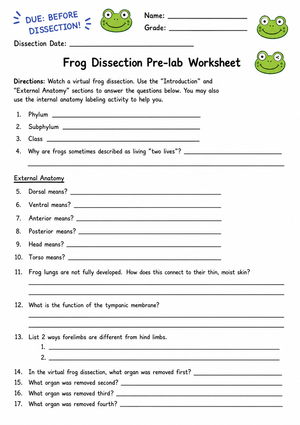
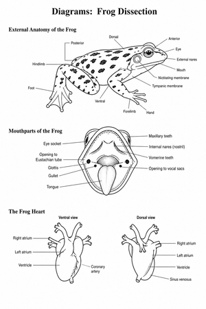
The sequence moves from simple identification to analytical reasoning. External anatomy comes first — students locate dorsal and ventral surfaces, identify the tympanic membrane, observe skin texture, and record digit count before any incision is made. That external stage is not preliminary busywork; it trains the systematic observation habits students need once the body cavity is open and organs are visible.
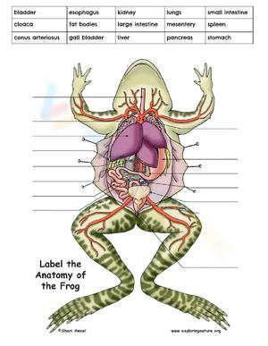
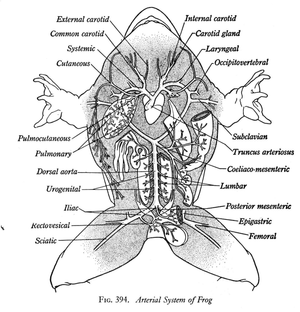
Internal anatomy work directs students through the major body systems in a logical order: digestive (liver, stomach, small intestine, large intestine, pancreas, gallbladder), circulatory (three-chambered heart, major vessels), respiratory (lungs, glottis, buccal floor), and reproductive (gonads, fat bodies). Organ-function matching tasks follow identification tasks — students first locate and label a structure, then explain its role in a separate section. Asking students to do both simultaneously while handling a specimen pushes cognitive load past the point where real learning happens, so the worksheets keep those tasks separate.
- Pre-lab vocabulary tasks: terms including tympanic membrane, cloaca, mesentery, fat bodies, and ventricle so students enter the lab with the language they need
- Safety checklist: glove use, scalpel and probe handling, specimen care, handwashing, and workspace cleanup — printed directly on the during-lab worksheet rather than left to verbal review alone
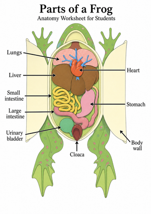
- Labeled anatomy diagrams: external and internal views, available in both blank and partially labeled versions
- Observation recording prompts: size, color, texture, and relative position of organs — written responses that go beyond naming
- Organ-function matching: a separate task that checks whether students understand what each structure does, not just what it is called
- Post-lab analysis questions: structure-to-function connections, system comparisons, and cross-species anatomy questions for students who move quickly
Fitting These Worksheets Into the Lab Period
The pre-lab worksheet works best assigned as homework the night before dissection day, or as a 10-minute warm-up before specimen trays arrive. Students who walk in having already matched vocabulary terms to definitions move through organ identification noticeably faster — they spend less time asking what a structure is called and more time recording accurate observations. That prior knowledge shows up directly in the quality of during-lab notes.
During the dissection itself, 9th grade frog dissection worksheets printable carry part of the classroom management load. The safety checklist at the top of the during-lab worksheet replaces the round of repeat questions that typically follows a verbal safety overview. The step-by-step observation sequence keeps groups moving at a similar pace, and the bounded tasks give students a clear stopping point at each stage. A practical technique that holds up in actual lab rooms: print the during-lab worksheet on a different color stock than the pre- and post-lab worksheets. When a group drifts off task, a single phrase — "you should be on the yellow sheet, internal organs section" — gets everyone back without interrupting other groups.
For standard 50-minute periods, the sequence splits cleanly: pre-lab vocabulary and external anatomy on day one, internal anatomy and organ-function matching on day two, post-lab analysis as a follow-up warm-up or assigned homework. Block periods of 80 to 90 minutes can accommodate the full sequence in one session, though most teachers find it useful to save the post-lab analysis worksheet for the next class — students write more carefully about structure and function after they have had time to process what they saw.
Errors That Show Up Consistently in Student Work
The liver catches almost every 9th grader off guard. When the body cavity opens, the liver's three dark-brown lobes fill most of the visual field, and students who have not internalized its location will label it as lungs, stomach, or simply leave it blank. A student who correctly identifies the liver on the pre-lab vocabulary worksheet will still misplace it on the dissection diagram because they are navigating the actual specimen orientation for the first time. A reference photograph on the during-lab worksheet — showing the liver in situ with a directional arrow — closes that gap faster than verbal correction during a live lab.
Intestine identification produces a consistent reversal error. Students expect the large intestine to be visibly wider than the small intestine, the way human anatomy diagrams present it. In a frog, that difference is much less pronounced. That expectation gap leads most students to switch the labels without second-guessing themselves. The observation prompt asking students to describe the relative width and coil pattern of each intestinal section gives them the specific language to sort it out rather than guessing from name alone.
The deeper pattern involves function rather than identification. Students describe organs in accurate sensory terms — "the lungs are small, pinkish sacs" — then write function answers that are vague or circular: "the lungs help the frog breathe." Post-lab questions that ask students to connect the lungs to buccal floor movement, or to explain why frog lungs are proportionally smaller than mammalian lungs, push past that surface-level answer and reveal what students actually understand about structure-function relationships.
Adjusting the Set for Different Learners
The 9th grade frog dissection worksheets printable include two diagram formats for the internal anatomy section. Blank diagrams ask students to label from memory and observation; partially labeled versions place some organ names and leave others blank. Both formats address the same anatomy content. The difference is in how much retrieval demand falls on the student during the live lab — when a student is simultaneously manipulating a probe, locating a structure, and trying to recall its name, a partially labeled diagram reduces that demand without removing the learning task entirely.
Word bank versions of the vocabulary and matching tasks give students access to correct terms without requiring full recall under lab conditions. These work especially well for students with limited prior biology exposure or those who are still building academic vocabulary in English. Students who don't need the word bank simply don't use it; its presence does not affect students working from complete recall.
For students who are not participating in hands-on specimen work — whether for personal, medical, or school policy reasons — the diagram and post-lab worksheets pair directly with a virtual dissection platform or a printed anatomy reference. Observation prompts describe what a structure looks like rather than how it feels under a probe, so students working from a screen or labeled photograph answer the same questions the rest of the class answers. Extension prompts for students who move quickly ask them to compare frog anatomy to a mammal's at the system level, or to hypothesize why a three-chambered heart functions adequately for an ectotherm while mammals require four chambers.
Standard Alignment
These worksheets align with NGSS performance expectation HS-LS1-2, which asks students to develop and use a model to illustrate the hierarchical organization of interacting systems within multicellular organisms. A dissected frog specimen is a physical model in the precise sense the standard intends — students observe structures nested within systems and can trace how those systems interact. The labeled diagram tasks produce the model; the post-lab analysis questions require students to use it to explain relationships among organ systems.
The disciplinary core idea LS1.A: Structure and Function underpins every organ-function task in the set. At the high school level, LS1.A expects students to explain how a structure's form contributes to the organism's overall function — not just to identify parts. The post-lab analysis questions are written specifically to meet that standard, asking for explanations of how form enables function rather than definitions alone.
Frequently Asked Questions
What should I do when a student's specimen is too degraded to identify all the organs?
This happens more often than teachers expect, especially with older preserved specimens where the liver may be discolored, the lungs collapsed, or the fat bodies dissolved. The partially labeled diagram version of the internal anatomy worksheet lets a student record what they can confirm visually and fill in remaining labels from the reference diagram. Students can also note in the observation section what was visible versus what wasn't — which is, in itself, a legitimate scientific observation worth documenting.
Can students who aren't dissecting still complete the worksheets meaningfully?
Yes. The pre-lab vocabulary worksheet and post-lab analysis questions require no specimen at all. For the identification and observation tasks, a virtual dissection platform provides the visual reference students need. The 9th grade frog dissection worksheets printable are written so that observation prompts describe what a structure looks like — students working from a screen can answer the same questions as students working with a physical specimen.
How do I grade these efficiently during a unit that already has multiple lab days?
Most teachers use the pre-lab worksheet as a completion check or brief readiness quiz, the during-lab observation notes as participation evidence, and the post-lab analysis questions as the primary written assessment. That approach spreads the evidence of learning across three manageable tasks rather than one high-stakes quiz, and it gives a fuller picture of student understanding. The organ-function matching section is particularly useful as a spot-check: if a student labels all organs correctly but cannot match them to functions, that is a specific instructional gap worth addressing before the unit assessment.
Clear All