


Views
Downloads
Printable Brain Anatomy Worksheet | Grade 7 Science
Paste this activity's link or code into your existing LMS (Google Classroom, Canvas, Teams, Schoology, Moodle, etc.).
Students can open and work on the activity right away, with no student login required.
You'll still be able to track student progress and results from your teacher account.
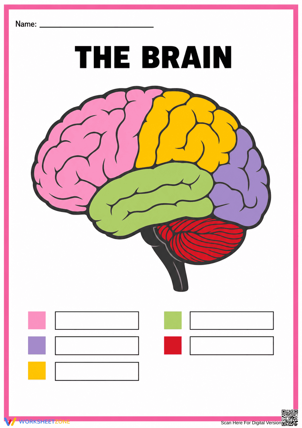
This Grade 7 brain anatomy worksheet enables students to identify and label the major regions of the human brain. By matching color-coded sections to their correct anatomical names, learners build a foundational understanding of the nervous system and how different lobes control specific bodily functions. It is an efficient tool for reinforcing biological structures.
At a Glance
- Grade: 7 · Subject: Science
- Standard:
MS-LS1-8— Identify how sensory receptors send messages to the brain.- Skill Focus: Brain anatomy and labeling
- Format: 1 page · 5 problems · Answer key included · PDF
- Best For: Independent practice or review
- Time: 10–15 minutes
This single-page resource features a clear, color-coded diagram of the human brain, highlighting five distinct anatomical regions. Students are tasked with completing five corresponding blank boxes using the provided color key. The visual scaffolding makes it easy for students to distinguish between the frontal, parietal, occipital, and temporal lobes, as well as the cerebellum. A complete answer key is included to ensure accurate grading and immediate student feedback.
Zero-Prep Workflow
- Print (1 minute): Simply download the PDF and print a class set. The design works perfectly in color or grayscale.
- Distribute (1 minute): Hand out the worksheet as a warm-up, exit ticket, or independent practice activity.
- Review (3 minutes): Use the included answer key to quickly check student responses or project it on the board for self-correction.
With under two minutes of total teacher prep time required, this activity is highly suitable for emergency sub plans or quick formative assessments during a busy science unit.
Standards Alignment
This activity aligns with MS-LS1-8: Gather and synthesize information that sensory receptors respond to stimuli by sending messages to the brain for immediate behavior or storage as memories. By identifying the specific regions of the brain, students establish the anatomical foundation necessary to understand where these sensory messages are processed. Both standard codes can be copied directly into lesson plans, IEP goals, or district curriculum mapping tools.
How to Use It
Teachers can integrate this labeling activity during direct instruction as a guided note-taking tool, or assign it after a lesson on the nervous system to check for understanding. For formative assessment, observe students to ensure they correctly match colors to lobes before writing. Expected completion time ranges from 10 to 15 minutes, making it a highly efficient instructional tool for middle school classrooms.
Who It's For
This worksheet is primarily designed for middle school life science students studying the human body systems. The strong visual cues and color-coding provide built-in differentiation, supporting English language learners and students who benefit from visual scaffolding. It pairs perfectly with a direct instruction lesson on the nervous system or an interactive anchor chart detailing the functions of each brain lobe.
Understanding the structural components of the nervous system is a critical step in mastering middle school life science concepts. This resource directly supports MS-LS1-8 by helping students identify how sensory receptors send messages to the brain. According to a recent EdReports 2024 analysis of effective science curricula, providing students with clear, color-coded anatomical diagrams significantly improves their ability to retain complex biological vocabulary and spatial relationships. When learners can visually map distinct regions like the frontal and occipital lobes, they build stronger cognitive frameworks for understanding how the body processes external stimuli. This targeted labeling exercise ensures students actively connect anatomical structures to physiological functions within the broader human body system. Integrating these visual scaffolds with rigorous scientific terminology fosters deeper comprehension and long-term retention of essential biological concepts.