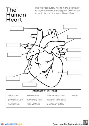
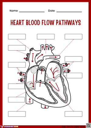
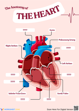
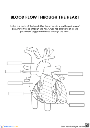
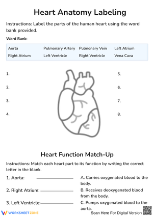
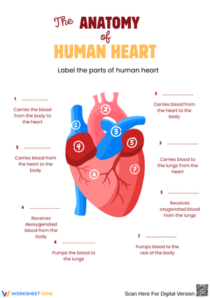
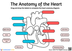
These labeling heart printable worksheets for 6th grade give science teachers a concrete way to check whether students have moved from memorizing vocabulary to actually understanding the spatial layout of cardiovascular anatomy. The set covers chambers, major vessels, internal structures, and blood-flow direction — the full scope of what middle school NGSS standards ask students to know about the heart. Each worksheet arrives as a print-ready PDF with clear diagrams and pointer lines sharp enough to survive the copier.
Anatomy Structures Covered Across the Set
The four chambers anchor each worksheet — right atrium, right ventricle, left atrium, and left ventricle. Students practice distinguishing between the atria's receiving function and the ventricles' pumping role, which trips up more students than the names themselves do. Several worksheets in the set include wall-thickness cues in the diagram so students notice that the left ventricle wall is substantially thicker than the right, connecting structure to function before the lesson even addresses it directly.
Beyond the chambers, each worksheet asks students to locate and label the major vessels: aorta, superior vena cava, inferior vena cava, pulmonary artery, and pulmonary veins. The septum appears across the set, and selected worksheets introduce all four valves — tricuspid, bicuspid, aortic, and pulmonary — with enough spatial context that students see what each valve is guarding rather than just matching a term to a pointer line.
Color-Coding Blood Flow Through the Heart
Several worksheets pair the labeling task with a color-coding exercise that makes the double-loop circulatory system visible on paper. Students use a blue colored pencil to trace deoxygenated blood from the body through the vena cava, into the right atrium, down to the right ventricle, and out through the pulmonary artery to the lungs. A red pencil marks the return path — oxygenated blood from the pulmonary veins into the left atrium, left ventricle, and out through the aorta. That two-path tracing forces students to hold the full circuit in mind at once rather than treating the left and right sides of the heart as unrelated structures.
This works particularly well when followed by a simple pulse lab. After students have traced the blood-flow paths on paper, measuring their resting and post-exercise heart rates gives the diagram a physiological anchor — the abstract arrows connect to something students can actually feel in their own wrists.
Common Misconceptions to Watch For and Correct
The most persistent error at this level is labeling the pulmonary artery as carrying oxygenated blood. Students are taught early that arteries carry oxygen-rich blood, and the pulmonary artery is the exception to that rule. Without a direct challenge to that assumption — which a blood-flow tracing task provides — students carry this error through the unit and onto the test. The worksheets surface it quickly, which is exactly when it's easiest to address.
A second error involves confusing atria and ventricles on the diagram itself. Students can recite "atria receive, ventricles pump" accurately but still flip the labels on a cross-section view, because the spatial positions feel counterintuitive — atria sit above the ventricles but are described as passive receiving chambers, and the mismatch between location and function causes second-guessing. Worksheets with long, clearly terminated pointer lines reduce that ambiguity without removing the challenge of the content.
Standard Alignment
These worksheets align with NGSS MS-LS1-3, which addresses structures and processes under the disciplinary core idea "From Molecules to Organisms." That standard asks students to construct explanations for how body systems interact to support life — not just identify parts, but explain why those parts are structured the way they are. Labeling heart printable worksheets for 6th grade address that requirement directly: students who can correctly locate the pulmonary artery and veins are positioned to explain why the cardiovascular and respiratory systems must work in tandem, which is the system-level reasoning the standard is actually measuring.
How to Build These Worksheets Into Your Lesson Plans
A blank heart diagram used as a bell-ringer on day one tells you immediately which students bring prior knowledge and which are starting from zero. Collect those without a grade, return them at the end of the unit, and let students compare what they labeled then against what they know now. That before-and-after move is low-effort formative assessment that also gives students a tangible record of their own growth across the unit.
For interactive science notebooks, print each worksheet at 80 percent scale. At that size, the diagram fits onto a standard composition notebook page without the edges overhanging, leaving margin space for blood-flow summaries or color-coded keys directly beside the diagram. Labeling heart printable worksheets for 6th grade also hold up well as dry-erase review tools — slide each worksheet into a clear page protector and students can label with a dry-erase marker, wipe it clean, and repeat. Running several short sessions across the days before a unit test takes advantage of spaced retrieval, which improves retention more reliably than a single long review block.
Differentiating These Worksheets Across Ability Levels
Students still building science vocabulary benefit from versions that include a word bank. The word bank removes the recall burden for the terms themselves so students can focus entirely on spatial placement — identifying where a structure is located rather than simultaneously trying to retrieve what it's called. Those are separate cognitive tasks, and combining them in one assessment often masks spatial reasoning ability in students who simply haven't consolidated the vocabulary yet.
For students ready for more challenge, remove the word bank and add a written-response prompt alongside the diagram: "Explain why the left ventricle wall is thicker than the right." That shifts the task from identification to reasoning and aligns more closely with what NGSS MS-LS1-3 is actually measuring. A further extension — asking students to draw and annotate blood-flow arrows on a blank heart outline without a pre-printed diagram — moves the work fully into application and works well as a task for students who finish early.
Frequently Asked Questions
What structures should a 6th grader be able to label on a heart diagram?
The core expectation at this level is the four chambers (right atrium, right ventricle, left atrium, left ventricle), the major vessels (aorta, superior and inferior vena cava, pulmonary artery, pulmonary veins), and the septum. The four valves — tricuspid, bicuspid, aortic, and pulmonary — are often introduced in 6th grade, but labeling requirements vary by district. Check your curriculum guide before deciding which structures to assess independently.
Can students reuse these worksheets for practice without printing extra copies?
Yes. Slide each worksheet into a clear page protector and students can label with a dry-erase marker, wipe it clean, and repeat. Running several short practice sessions across the days before a unit test — rather than one long review — takes advantage of spaced retrieval, which produces stronger long-term retention than massed practice does.
Do the worksheets come with answer keys?
Labeling heart printable worksheets for 6th grade in this set include a fully labeled answer key for each diagram. Teachers use them for quick grading or set them up as self-checking stations where students verify their own work immediately after completing the labeling task — which gives students faster feedback than waiting for a returned assignment.









 Interactive Worksheet Maker
 Quiz Maker
 Word Search Maker
 Crossword Puzzle Maker
 Word Scramble Generator
 Sentence Scramble Generator
 Handwriting Worksheet Generator
 Name Tracing Generator
 Fill In The Blank Generator




 BACK TOSCHOOL