




























Complete Pig Anatomy CER Worksheet | Grades 7–12 Science
Paste this activity's link or code into your existing LMS (Google Classroom, Canvas, Teams, Schoology, Moodle, etc.).
Students can open and work on the activity right away, with no student login required.
You'll still be able to track student progress and results from your teacher account.
This fetal pig anatomy worksheet guides secondary biology students through scientific explanation using the Claim-Evidence-Reasoning (CER) framework. Students examine heart diagrams, evaluate blood oxygen saturation levels, and construct evidence-based arguments about fetal circulation shunts and organ system interactions. It bridges anatomical observation with analytical scientific writing across 22 targeted tasks.
At a Glance
- Grade: Grades 7–12 · Subject: Science
- Standard: Structure and Function — Construct explanations linking anatomical structures to circulatory and respiratory system functions
- Skill Focus: Claim-Evidence-Reasoning in fetal pig anatomy
- Format: 4 pages · 22 problems · Answer key included · PDF
- Best For: Biology dissection labs and data analysis
- Time: 45–60 minutes
The 4-page activity packet features a labeled diagram of the fetal heart, a blood oxygen saturation data table, and five recorded dissection observations. Students progress through anatomical identification, quantitative data interpretation, and multi-step written arguments. The complete 4-page answer key provides sample student claims, cited evidence, and physiological reasoning for immediate grading.
- Guided practice (Part A, 7 items): Students identify labeled structures in a fetal heart diagram and answer targeted multiple-choice questions about the foramen ovale and ductus arteriosus bypasses.
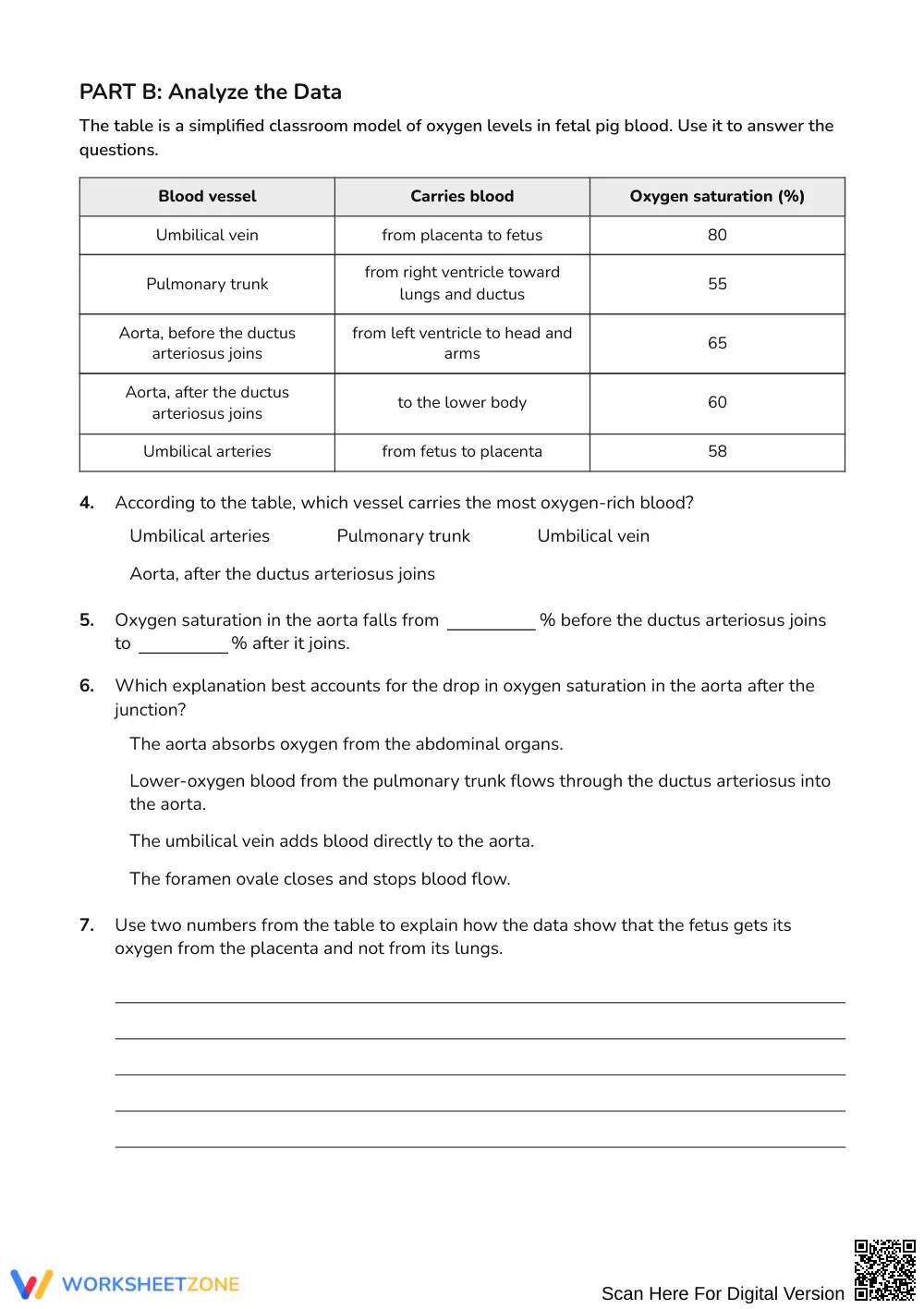
- Supported practice (Part B, 5 items): Students interpret quantitative blood oxygen saturation data across major vessels to trace maternal-fetal gas exchange.
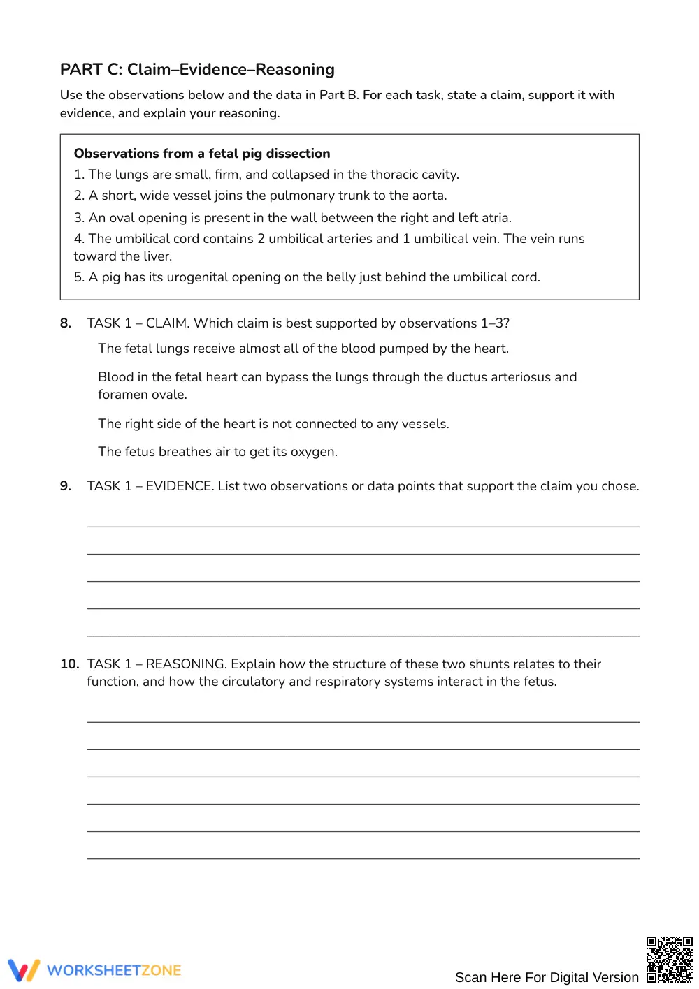
- Independent practice (Part C, 10 items): Students formulate claims, cite anatomical data, and compose reasoning to evaluate physiological changes after birth and determine specimen sex.
This sequence follows a gradual-release instructional model that moves students from structural identification to independent synthesis.
This activity aligns with secondary life science standards emphasizing how organ systems interact and how specialized body structures perform specific life functions. The tasks emphasize analyzing data to construct scientific explanations. These alignment points integrate cleanly into biology curriculum maps and laboratory performance assessments.
Implement this worksheet immediately after introducing fetal circulation or as an analytical companion during a fetal pig dissection unit. For a 50-minute lab period, have students examine the heart model before interpreting the saturation table. As a formative assessment check, monitor Part C Task 1 to confirm students explain why blood bypasses collapsed lungs before birth.
This resource supports general biology, advanced life science, and introductory anatomy courses. Scaffolded sentence prompts assist developing writers in structuring evidence, while the comparative data table challenges advanced learners. Pair this packet with a mammalian heart dissection or an organ system anchor chart.
The Claim-Evidence-Reasoning framework applied in this worksheet grounds scientific inquiry in observable data and biological principles. According to Fisher & Frey (2014), structured writing scaffolds and gradual release models significantly increase student mastery of complex domain-specific discourse, especially when synthesizing quantitative evidence with visual models. In secondary biology classrooms, connecting structural anatomy directly to physiological mechanisms remains a frequent obstacle for developing learners. By prompting students to evaluate circulation data, identify maternal-fetal oxygen pathways, and justify why fetal cardiovascular shunts close after birth, this resource reinforces deep conceptual understanding rather than passive memorization. The multi-tiered sequence of labeling, data analysis, and argumentation ensures that students practice authentic disciplinary literacy. Teachers can extract this instructional framework directly for lab evaluation, portfolio documentation, or standards-based progress tracking.


