


Views
Downloads
Printable Mitosis Stages Worksheet | Grade 10 Biology
Paste this activity's link or code into your existing LMS (Google Classroom, Canvas, Teams, Schoology, Moodle, etc.).
Students can open and work on the activity right away, with no student login required.
You'll still be able to track student progress and results from your teacher account.
This Grade 10 biology worksheet provides a comprehensive review of the mitotic cycle, helping students master cell division. By analyzing visual representations of animal and plant cells, learners develop a concrete understanding of how genetic material replicates and segregates to maintain life.
At a Glance
- Grade: 10 · Subject: Biology
- Standard:
HS-LS1-4— Use models to illustrate cellular division- Skill Focus: Mitosis Phase Identification
- Format: 3 pages · 8 problems · Answer key included · PDF
- Best For: Formative assessment and review
- Time: 30–45 minutes
The 3-page PDF contains 8 multi-part questions designed to reinforce cellular biology concepts. It includes detailed animal cell diagrams for labeling, microscopic-style drawings of plant cells in an onion root tip, and a creative coloring task to track chromosome movement. A full answer key is provided to ensure accurate grading and immediate student feedback.
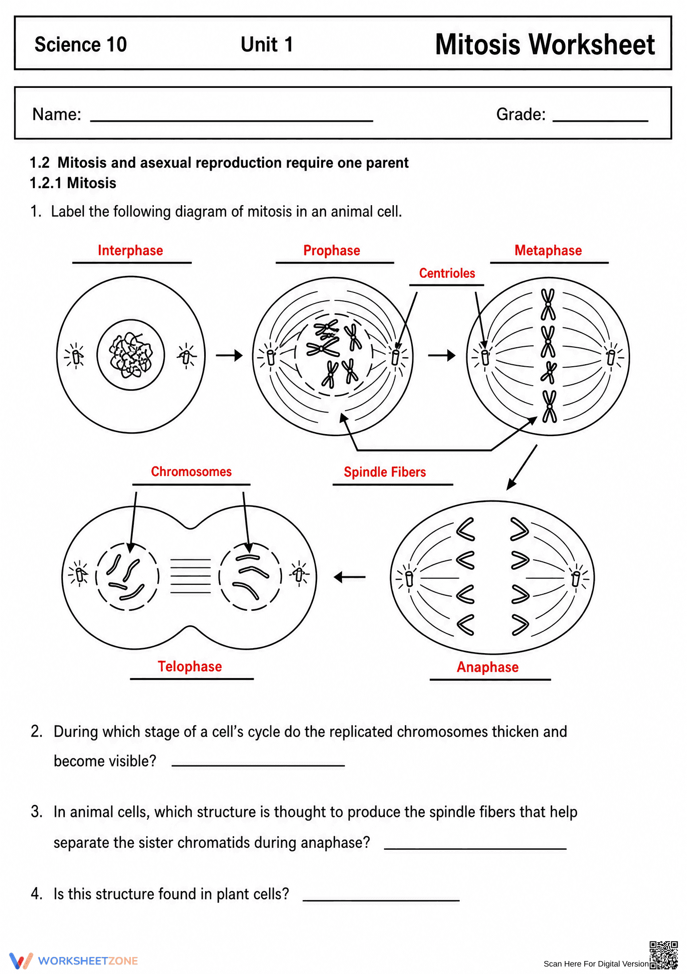
- Guided practice: Students begin by labeling a complete animal cell mitosis diagram with provided terms, identifying 5 key phases and structures like spindle fibers.
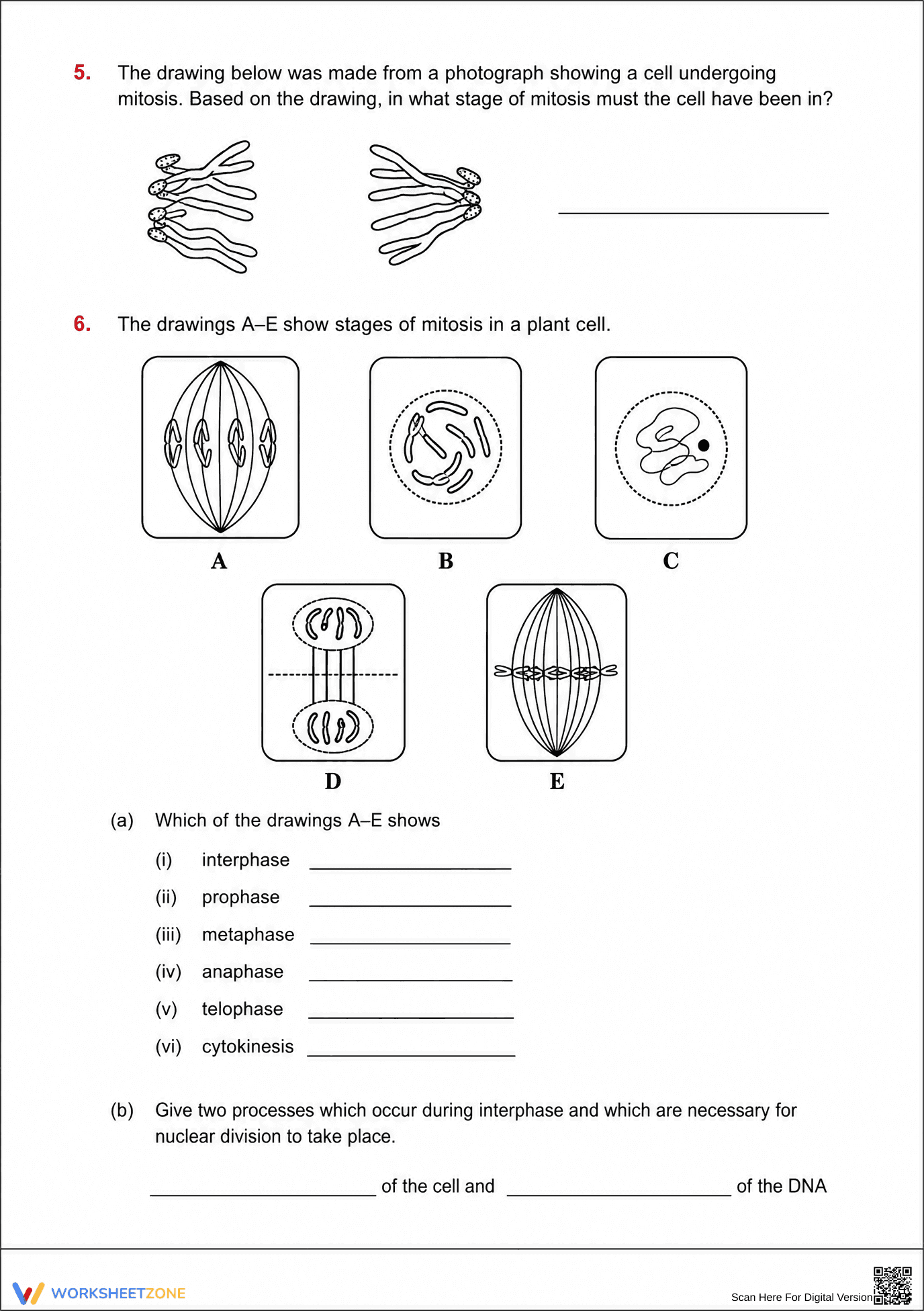
- Supported practice: Learners match specific cellular characteristics to plant cell drawings, identifying the progression from interphase through cytokinesis in 6 distinct steps.
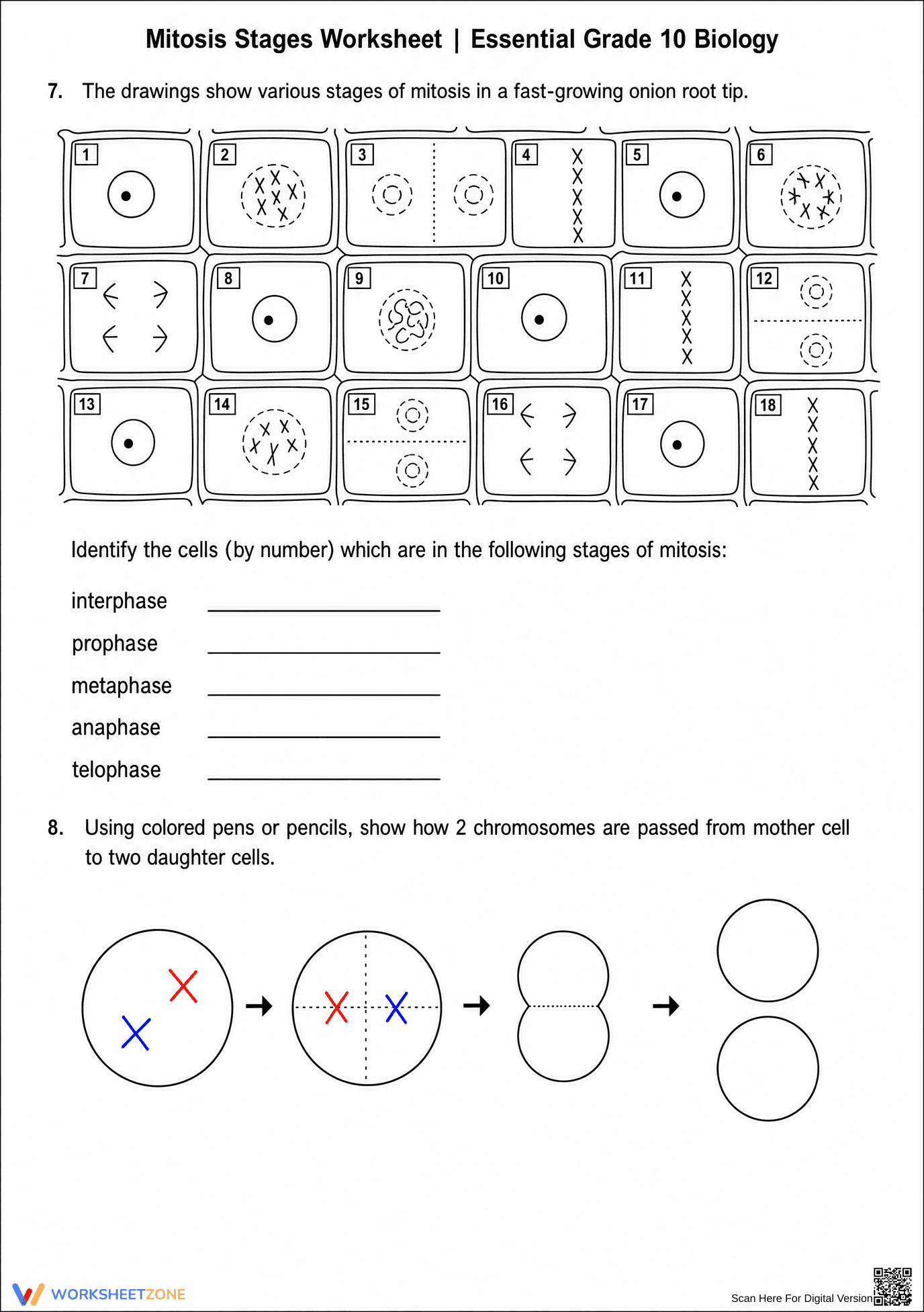
- Independent practice: The final tasks require students to identify stages in a complex 18-cell tissue sample and model chromosome distribution through independent drawing.
This sequence follows a gradual-release model, moving from simple identification to complex visual analysis of cellular structures.
This resource aligns with HS-LS1-4: Use a model to illustrate the role of cellular division (mitosis) and differentiation in producing and maintaining complex organisms. It specifically addresses the structural changes in chromosomes and organelles during the M-phase. Both standard codes can be copied directly into lesson plans, IEP goals, or district curriculum mapping tools.
Use this as a formative assessment after a direct instruction lecture on the cell cycle, or assign it as a collaborative lab activity where students compare the worksheet diagrams to actual slides under a microscope. Expect students to spend approximately 30 to 45 minutes completing all 8 sections. While circulating, observe if students can accurately distinguish between telophase and cytokinesis in the plant cell models during the identification phase.
Designed for Grade 9 and 10 biology students, including those in honors or general science tracks. It is particularly helpful for visual learners who require concrete representations of abstract biological processes. Pair this worksheet with a 3D cell model or a digital animation of the mitotic spindle for maximum instructional impact.
According to the RAND AIRS 2024 report on secondary science instruction, the use of high-quality visual models is critical for student mastery of microscopic biological processes. This worksheet addresses the HS-LS1-4 standard by requiring students to interpret and create models of mitosis, a fundamental skill in cellular biology. Research suggests that scaffolded diagram labeling, as seen in these 8 tasks, significantly improves long-term retention of complex terminology. By transitioning from simple animal cell diagrams to complex onion root tip identification, the resource ensures students can apply their knowledge to varied biological contexts. This 3-page document provides the necessary rigor for high school biology while maintaining accessibility through clear visual cues and structured prompts, making it a reliable tool for meeting state and national life science benchmarks.