


Views
Downloads
Mitosis Phases Worksheet | Grade 9-12 Essential
Paste this activity's link or code into your existing LMS (Google Classroom, Canvas, Teams, Schoology, Moodle, etc.).
Students can open and work on the activity right away, with no student login required.
You'll still be able to track student progress and results from your teacher account.
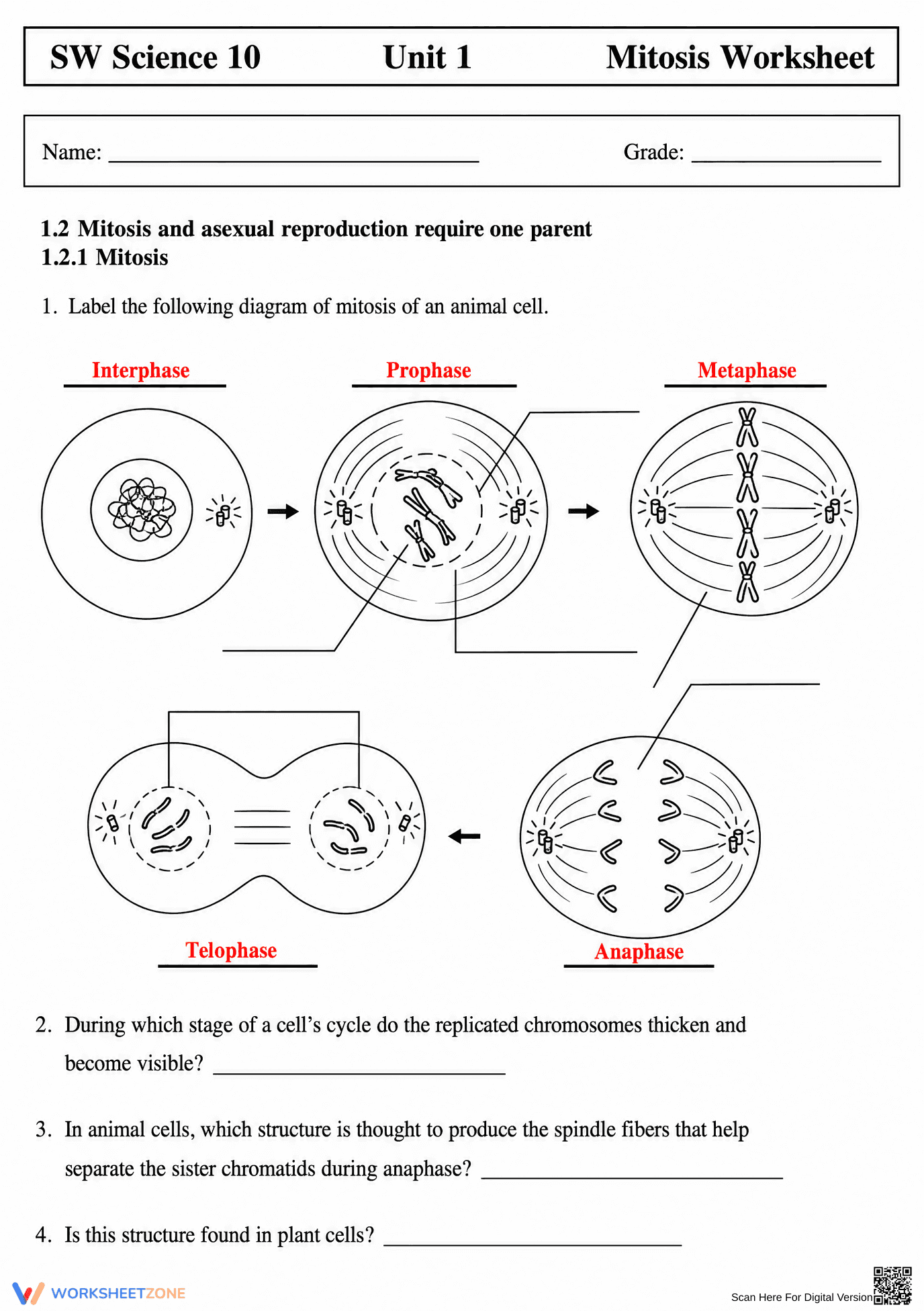
This comprehensive biology resource provides students with a structured way to master the complex stages of cellular division. By moving from simple labeling to identifying real-world microscopic views, learners develop a deep conceptual understanding of how genetic material is organized and partitioned. Students will successfully differentiate between prophase, metaphase, anaphase, and telophase across multiple cell types.
At a Glance
- Grade: 9–12 · Subject: Biology
- Standard:
HS-LS1-4— Use models to illustrate how cellular division produces and maintains complex organisms- Skill Focus: Mitosis phase identification
- Format: 3 pages · 32 tasks · Answer key included · PDF
- Best For: High school biology unit review
- Time: 45–60 minutes
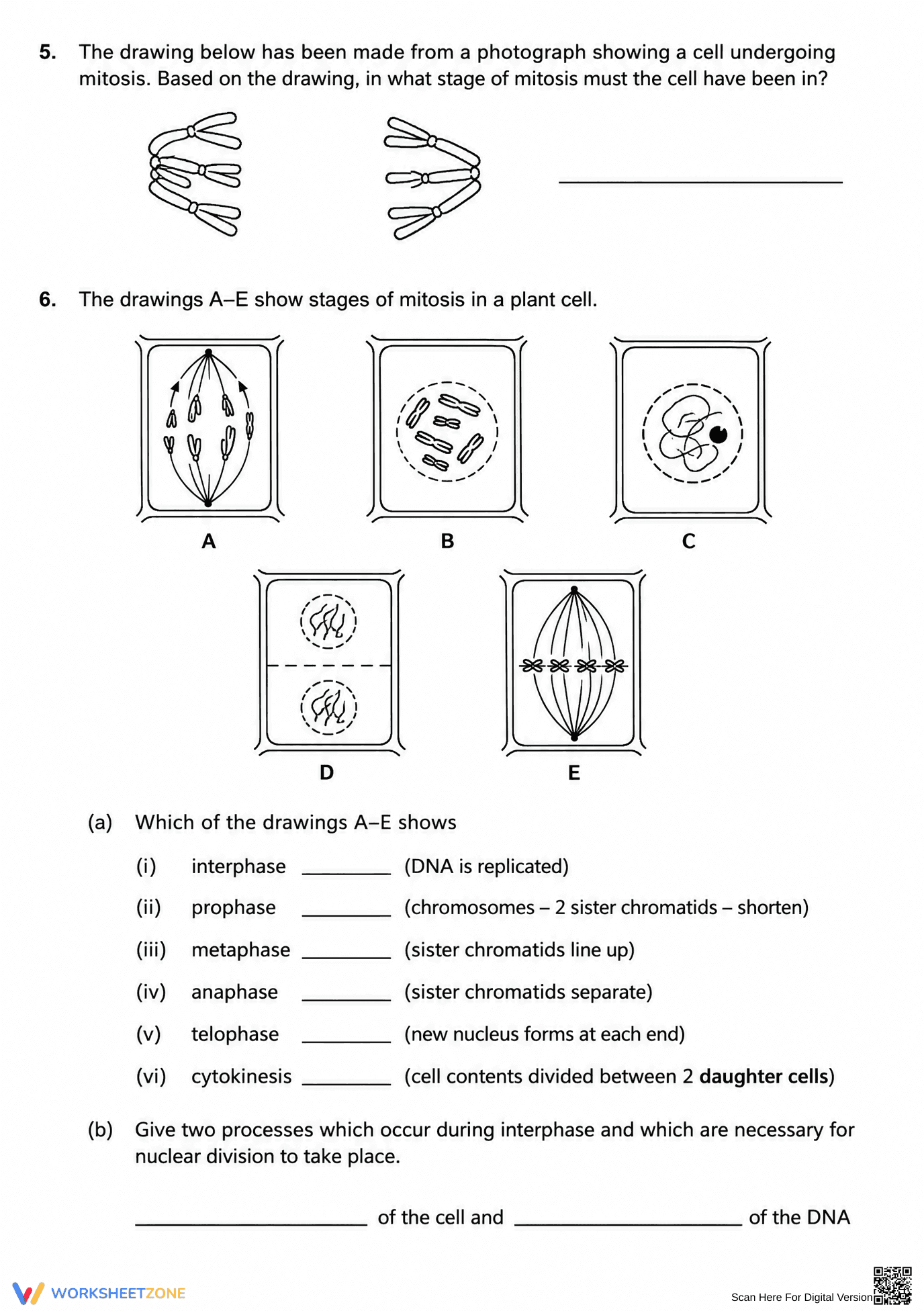
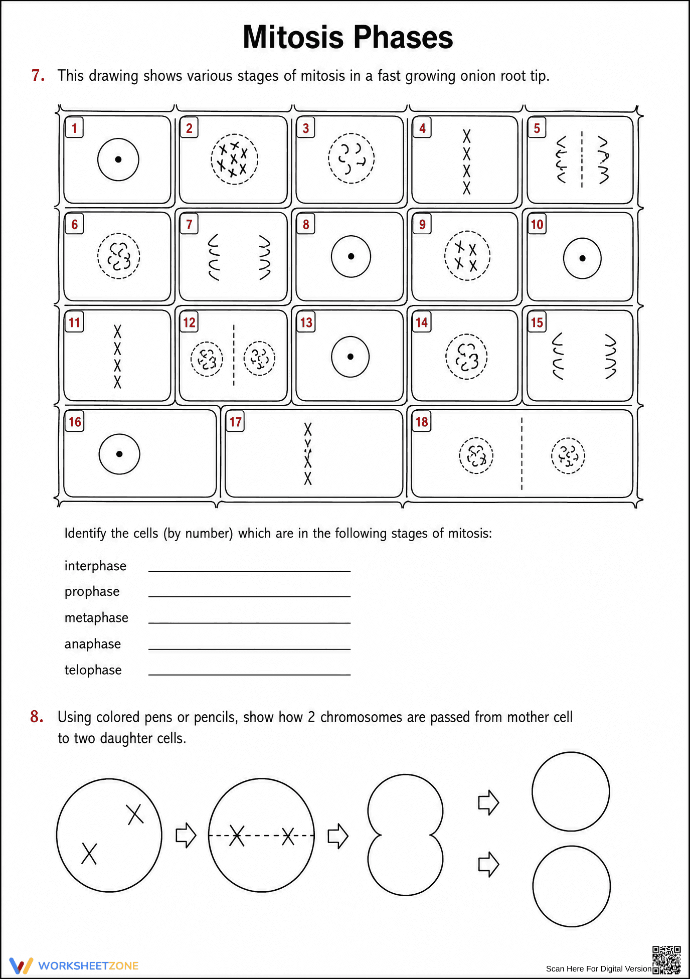
This 3-page PDF includes a detailed animal cell diagram for initial labeling, a plant cell comparison section, and a high-rigor identification grid featuring 18 different onion root tip cells. The worksheet concludes with a modeling task where students use colored pens to track chromosome passage from a mother cell to two daughter cells. A full answer key is provided to facilitate quick grading.
This resource is designed for immediate classroom implementation with a total teacher prep time of less than two minutes. First, print the three-page set for your class (1 minute). Second, distribute the packets as a standalone summative assessment or a guided laboratory supplement (1 minute). Finally, use the included answer key to review the 18-cell identification grid as a whole-class activity to clear up common misconceptions (15 minutes).
The primary focus is HS-LS1-4: "Use a model to illustrate the role of cellular division (mitosis) and differentiation in producing and maintaining complex organisms." This worksheet specifically addresses the modeling component by requiring students to visualize and draw the physical arrangement of chromosomes. Both standard codes can be copied directly into lesson plans, IEP goals, or district curriculum mapping tools.
Use this worksheet as a mid-unit formative assessment after students have been introduced to the cell cycle. It works exceptionally well as a "dry lab" before students view actual onion root tip slides under a microscope. During the activity, observe if students can distinguish between the alignment of chromosomes in metaphase versus their separation in anaphase; this is a critical indicator of readiness for advanced genetics.
This resource is tailored for Grade 9-12 Biology students, including those in Honors tracks who need to reinforce the visual identification of mitotic phases. It provides enough scaffolding for general education students while offering the complexity required for advanced learners. Pair this with a digital animation of the cell cycle or a physical bead-modeling activity for a multi-modal learning experience.
According to research by Fisher & Frey (2014) on the gradual release of responsibility, providing students with multiple visual representations—such as the animal, plant, and onion root tip diagrams found here—is essential for moving from guided instruction to independent mastery. This worksheet aligns with the HS-LS1-4 standard by requiring students to identify and model the specific structural changes that occur during the M-phase of the cell cycle. By analyzing 18 distinct cell images, students engage in the high-repetition practice necessary to internalize the nuances of cellular biology. Data from NAEP science assessments suggest that students who can successfully model biological processes show higher retention rates of complex systems. This printable resource ensures that every learner has a concrete reference point for the mechanics of asexual reproduction and tissue growth.