


0
Views
0
Downloads
0.0
0
Save
0 Likes
Hamstring Muscles Anatomy Diagram
0 Views
0 Downloads
Paste this activity's link or code into your existing LMS (Google Classroom, Canvas, Teams, Schoology, Moodle, etc.).
Students can open and work on the activity right away, with no student login required.
You'll still be able to track student progress and results from your teacher account.
Information
Description
What It Is:
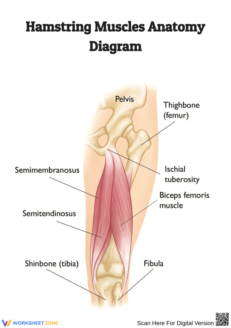
This hamstring muscles anatomy diagram shows the key muscles of the posterior thigh—semimembranosus, semitendinosus, and biceps femoris—along with related structures such as the pelvis, femur, tibia, fibula, and ischial tuberosity. It provides a clear, labeled overview of the hamstring region.
Why Use It:
This labeled diagram helps students, athletes, and medical learners visualize hamstring structure and attachment points. It’s useful for understanding movement, strength training, rehabilitation, and sports injury prevention.
How to Use It:
• Study the labeled muscles to learn their locations and functions.
• Use it as a reference for anatomy lessons, physiotherapy, or sports science.
• Pair with worksheets on labeling hamstrings or tracing their functions in movement.
• Apply in fitness or rehabilitation contexts to understand muscle engagement.
Grade Suitability:
Best suited for high school to higher education.
• High school biology and anatomy students.
• College and medical school learners.
• Sports science and physiotherapy training.
Target Users:
Teachers, anatomy instructors, physiotherapy educators, medical students, and athletes studying hamstring muscle function.