

























Views
Downloads

Printable Virus Life Cycle Diagram | Grade 9-12 Science
Paste this activity's link or code into your existing LMS (Google Classroom, Canvas, Teams, Schoology, Moodle, etc.).
Students can open and work on the activity right away, with no student login required.
You'll still be able to track student progress and results from your teacher account.
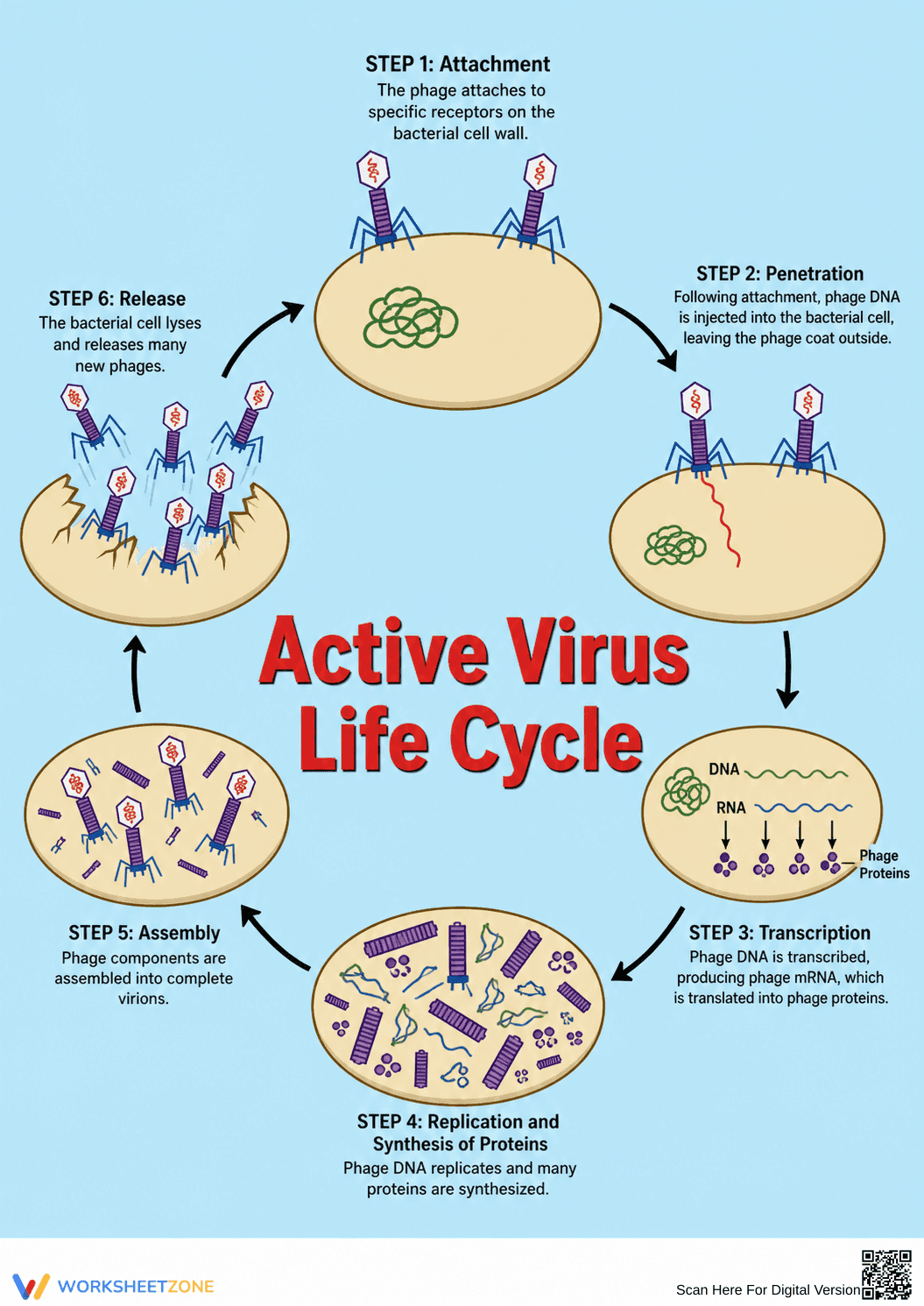
This high school biology reference diagram helps students visualize the active virus life cycle, specifically focusing on bacteriophages. By breaking down the lytic cycle into six distinct stages, learners can easily grasp how viruses hijack host cells to replicate and synthesize proteins.
At a Glance
- Grade: 9 · Subject: Biology
- Standard:
HS-LS1-1— Explain how DNA determines protein structure and cellular functions.- Skill Focus: Analyzing viral replication stages
- Format: 1 page · 6 stages · No answer key needed · PDF
- Best For: Visual reference and study guide
- Time: 5–10 minutes
This single-page visual guide features a circular diagram detailing six critical steps of bacteriophage infection: attachment, penetration, transcription, replication, assembly, and release. Each phase includes an illustration of the virus interacting with a bacterial host cell, paired with concise text defining key terms like phage DNA and lysis. It serves as a complete study aid.
Zero-Prep Workflow
- Print (1 minute): Download the PDF and print copies for student binders or project them directly onto a smartboard.
- Distribute (1 minute): Hand out the diagram as a companion resource during your lecture on viral mechanisms.
- Review (3 minutes): Walk through the six stages together, highlighting how the virus uses the host's machinery to synthesize proteins.
With zero teacher setup required, this resource is highly effective for immediate classroom use or as a reliable addition to a substitute teacher plan.
Standards Alignment
This resource aligns with HS-LS1-1: "Construct an explanation based on evidence for how the structure of DNA determines the structure of proteins which carry out the essential functions of life through systems of specialized cells." It directly supports this by illustrating how viral DNA interrupts and utilizes these cellular systems. Both standard codes can be copied directly into lesson plans, IEP goals, or district curriculum mapping tools.
How to Use It
Introduce this diagram during direct instruction when transitioning to pathogens. Students can keep it on their desks as a quick-reference guide while completing assignments. For a quick formative assessment, cover the text labels on a projected version and ask students to verbally explain a specific stage based solely on visual cues. Expect students to spend 5 to 10 minutes reviewing the cycle.
Who It's For
This diagram is designed for high school biology students, though it can be adapted for advanced middle school life science classes. The clear visual scaffolding supports English Language Learners and visual learners who might struggle with dense textbook descriptions of viral replication. Pair this reference sheet with a blank graphic organizer to encourage active recall and reinforce the sequence of events.
Understanding complex biological processes requires strong visual supports that reduce cognitive load and clarify microscopic interactions. When students analyze the active virus life cycle, they must explain how DNA determines protein structure and cellular functions, a core requirement of HS-LS1-1. According to a 2024 report by EdReports, integrating clear, sequenced diagrams into science instruction significantly improves student retention of microscopic mechanisms that cannot be observed directly in a standard classroom laboratory. By providing a step-by-step visual breakdown of bacteriophage infection—from initial attachment to final cellular lysis—educators can help learners connect abstract vocabulary like transcription and protein synthesis to concrete visual models. This instructional approach not only builds foundational biology knowledge but also strengthens overall scientific literacy, ensuring students can accurately interpret, analyze, and communicate complex cellular interactions during assessments and future scientific coursework.


