




























Views
Downloads

Printable Integumentary System Worksheet | Grade 7 Science

Paste this activity's link or code into your existing LMS (Google Classroom, Canvas, Teams, Schoology, Moodle, etc.).
Students can open and work on the activity right away, with no student login required.
You'll still be able to track student progress and results from your teacher account.
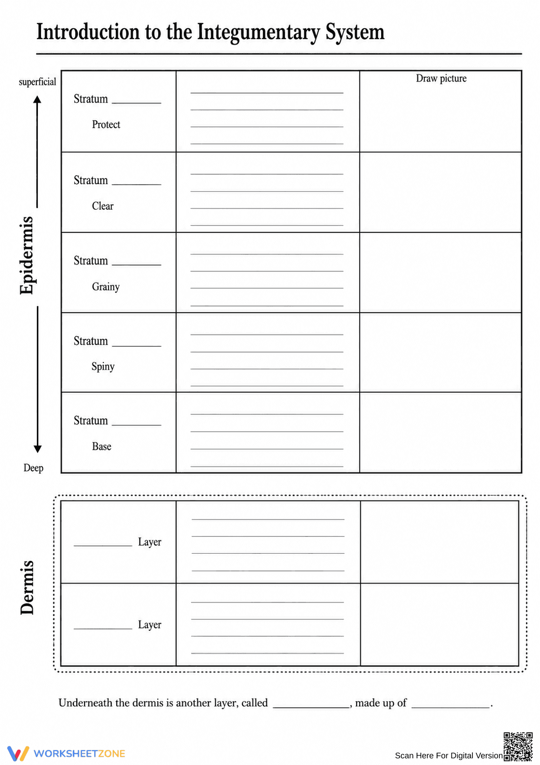
This printable integumentary system worksheet helps students identify and describe the specific layers of the skin. By completing this structured graphic organizer, learners will master the five strata of the epidermis and the two layers of the dermis, reinforcing their understanding of human body subsystems and anatomical organization.
At a Glance
- Grade: 7 · Subject: Science
- Standard:
MS-LS1-3— Describe how the body is a system of interacting subsystems- Skill Focus: Identifying skin layers
- Format: 1 page · 8 problems · Answer key included · PDF
- Best For: Independent practice or review
- Time: 15–20 minutes
Inside this resource, you will find a single-page graphic organizer that breaks down the integumentary system from superficial to deep layers. Students name each specific stratum, write a brief description of its function, and draw a corresponding picture to visualize the cellular structure. A complete answer key is provided.
Designed for immediate classroom use with a zero-prep workflow:
- Print (1 minute): Simply download the PDF and print a class set. The black-and-white design is ink-friendly.
- Distribute (1 minute): Hand out the graphic organizer after your initial lesson. Instructions are self-explanatory.
- Review (3 minutes): Use the included answer key to quickly check student work or project it for self-grading.
Total teacher prep time is under two minutes, making this an excellent option for sub plans.
This resource is aligned to the Next Generation Science Standard MS-LS1-3: Use argument supported by evidence for how the body is a system of interacting subsystems composed of groups of cells. By detailing the specific layers of the epidermis and dermis, students build a foundational understanding of how specialized cellular structures form the skin subsystem. Both standard codes can be copied directly into lesson plans, IEP goals, or district curriculum mapping tools.
This worksheet works well as independent practice immediately following direct instruction. Students can use textbooks to fill in descriptions and sketch layers. Alternatively, it serves as an effective review guide before an exam. As a formative assessment tip, observe students while they draw; this visual component reveals if they grasp structural differences between the epidermal layers. Expect completion in 15 to 20 minutes.
This resource is primarily designed for middle school science and early high school biology students studying human anatomy. The inclusion of a drawing column provides built-in differentiation for visual learners who might struggle with rote memorization of anatomical terms. It pairs perfectly with a 3D skin model demonstration or an introductory slide deck on human body systems, giving students a structured place to record their observations.
Integrating structured graphic organizers into anatomy instruction significantly improves student retention of complex biological systems. According to a ScienceDirect TpT Analysis, resources that combine linguistic recall with visual representation—such as drawing cellular structures alongside their scientific names—enhance cognitive processing and long-term memory. This worksheet directly supports MS-LS1-3 by requiring students to describe how the body is a system of interacting subsystems. When learners actively map out the superficial to deep layers of the integumentary system, they move beyond simple memorization and begin to understand the functional relationships between different tissue types. Providing a dedicated space for both written descriptions and visual sketches ensures that multiple learning modalities are engaged simultaneously, leading to deeper comprehension of human body organization and cellular specialization.


