


Views
Downloads
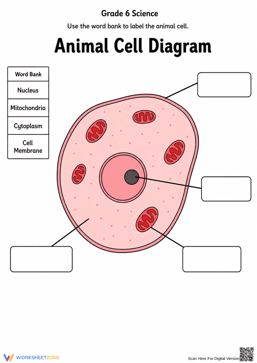
Essential Animal Cell Diagram Worksheet | Grade 6
Paste this activity's link or code into your existing LMS (Google Classroom, Canvas, Teams, Schoology, Moodle, etc.).
Students can open and work on the activity right away, with no student login required.
You'll still be able to track student progress and results from your teacher account.
This Grade 6 science worksheet provides a clear, visual method for students to identify the fundamental components of an animal cell. By matching terms from a word bank to a high-quality diagram, learners solidify their understanding of cellular anatomy and the spatial relationships between organelles. It serves as a foundational tool for introductory biology units.
At a Glance
- Grade: 6 · Subject: Science
- Standard:
MS-LS1-2— Develop and use a model to describe the function of a cell- Skill Focus: Organelle Identification
- Format: 1 page · 4 problems · Answer key included · PDF
- Best For: Quick formative assessment or bell ringer
- Time: 5–10 minutes
The resource features a single-page layout centered on a detailed, color-coded animal cell illustration. It includes a dedicated word bank containing four essential biological terms: Nucleus, Mitochondria, Cytoplasm, and Cell Membrane. Four distinct call-out boxes are strategically placed with leader lines pointing to the corresponding structures, ensuring students can clearly distinguish between the cell's internal and external boundaries.
This worksheet is designed for immediate classroom implementation with a total teacher prep time of under 2 minutes. First, print the single-page PDF for your class size. Second, distribute the sheets as a "do-now" activity or during the "We Do" phase of a lesson. Third, review the answers collectively using the diagram as a visual anchor on a projector. Its self-explanatory nature makes it an ideal candidate for emergency sub plans or independent station work.
This resource is aligned with MS-LS1-2: "Develop and use a model to describe the function of a cell as a whole and ways parts of cells contribute to the function." While focused on identification, it provides the necessary vocabulary for students to later explain how these parts interact. This standard code can be copied directly into lesson plans, IEP goals, or district curriculum mapping tools.
Use this worksheet as a formative assessment immediately following a direct instruction session on cell theory. As students work, circulate to observe if they can distinguish the nucleus from the mitochondria based on visual cues. It also functions effectively as a post-unit review. Expect students to complete the labeling in approximately 5 to 10 minutes, depending on prior knowledge.
This activity is tailored for middle school students, specifically those in Grade 6, who are encountering life science concepts for the first time. The visual nature of the diagram supports English Language Learners and students with processing needs by providing a word bank scaffold. It pairs naturally with a microscope lab or an interactive anchor chart showing cell functions.
The use of visual models like this animal cell diagram is supported by the research of Fisher & Frey (2014), who emphasize the importance of gradual release of responsibility through scaffolded visual aids. By providing a word bank alongside a clear spatial model, this worksheet reduces cognitive load, allowing students to focus on the specific characteristics of the MS-LS1-2 standard. Identifying the nucleus, mitochondria, cytoplasm, and cell membrane is a critical prerequisite for understanding complex biological systems. According to the RAND AIRS 2024 analysis of instructional materials, high-quality visual representations are essential for long-term retention of abstract scientific concepts in middle school learners. This resource provides a structured entry point for students to master the foundational vocabulary required for higher-level biology and life science curriculum standards across various state frameworks.