


0
Views
0
Downloads
0.0
0
Save
0 Likes
Cell Anatomy Worksheet: Exploring Cell Structure
0 Views
0 Downloads
Paste this activity's link or code into your existing LMS (Google Classroom, Canvas, Teams, Schoology, Moodle, etc.).
Students can open and work on the activity right away, with no student login required.
You'll still be able to track student progress and results from your teacher account.
Information
Description
What It Is:
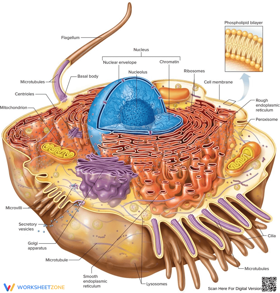
This is a detailed diagram of a eukaryotic cell. It shows a cross-section view, revealing internal organelles like the nucleus, nucleolus, chromatin, ribosomes, rough endoplasmic reticulum, smooth endoplasmic reticulum, Golgi apparatus, mitochondria, lysosomes, peroxisome, centrioles, microtubules, microvilli, cilia, flagellum, and basal body. A zoomed-in section illustrates the phospholipid bilayer structure of the cell membrane. Each part of the cell is labeled with an arrow pointing to it.
Grade Level Suitability:
This diagram is suitable for high school biology (Grades 9-12) and introductory college biology courses. The complexity of the cell structure and the level of detail in the labeling are appropriate for students who are learning about cell biology at a more advanced level.
Why Use It:
This diagram is useful for visually understanding the structure and organization of a eukaryotic cell. It helps students learn the names and locations of different organelles within the cell. It also helps to understand the cell membrane structure. It promotes visual learning and reinforces concepts taught in textbooks and lectures.
How to Use It:
Use this diagram as a reference while studying cell biology. You can use it to identify and label the different parts of the cell. It can also be used as a study aid to quiz yourself on the functions of each organelle. The diagram can be used for presentations, reports, or as a visual aid for teaching.
Target Users:
This diagram is beneficial for high school and college students studying biology, as well as teachers who are teaching cell biology. It is also useful for anyone interested in learning more about the structure of eukaryotic cells.