


0
Views
0
Downloads
0.0
0
Save
0 Likes
Cytology: Cytoplasm Cell Worksheet for Students
0 Views
0 Downloads
Paste this activity's link or code into your existing LMS (Google Classroom, Canvas, Teams, Schoology, Moodle, etc.).
Students can open and work on the activity right away, with no student login required.
You'll still be able to track student progress and results from your teacher account.
Information
Description
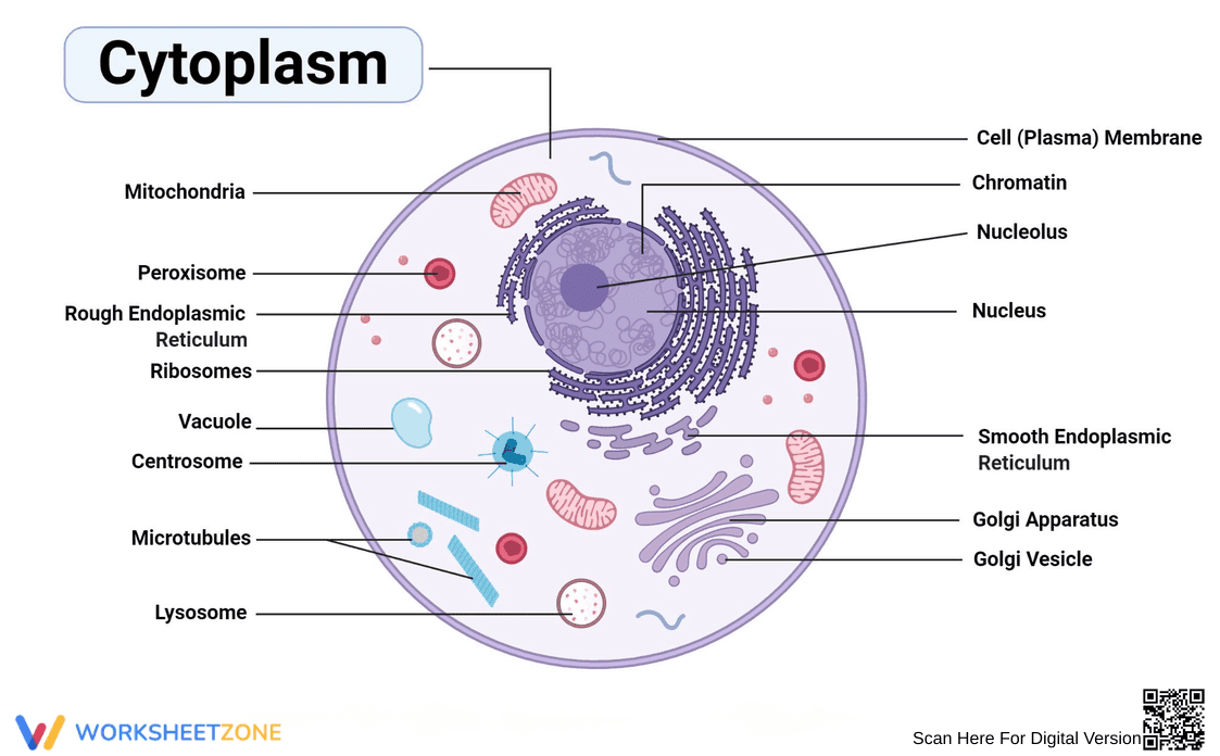
What It Is:
This is an educational worksheet featuring a labeled diagram of an animal cell, specifically focusing on the Cytoplasm. It identifies key organelles within the cell, including the Mitochondria, Peroxisome, Rough Endoplasmic Reticulum with Ribosomes, Vacuole, Centrosome, Microtubules, Lysosome, Cell (Plasma) Membrane, Chromatin, Nucleolus, Nucleus, Smooth Endoplasmic Reticulum, Golgi Apparatus, and Golgi Vesicle. The image provides a visual representation of cell structure.
Grade Level Suitability:
This worksheet is suitable for middle school (grades 6-8) and early high school (grades 9-10) biology classes. The labeled diagram provides a clear introduction to cell structure and organelles, making it appropriate for students learning about cells for the first time or reviewing basic cell biology concepts.
Why Use It:
This worksheet helps students visualize the components of an animal cell and understand the location and names of various organelles within the cytoplasm. It promotes visual learning and reinforces vocabulary related to cell biology. It also serves as a reference guide for identifying organelles.
How to Use It:
The worksheet can be used as a visual aid during a lesson on cell structure. Students can use it to identify and label cell organelles. It can also be used for review or as a study guide. Teachers can use it as a basis for quizzes or assignments requiring students to identify and describe the function of different organelles.
Target Users:
The target users are middle and high school students studying biology, as well as teachers who need a clear and concise diagram of an animal cell for their lessons. It is also helpful for students who are visual learners.