


0
Views
0
Downloads
0.0
0
Save
0 Likes
Facial Muscles: A Detailed Anatomy
0 Views
0 Downloads
Paste this activity's link or code into your existing LMS (Google Classroom, Canvas, Teams, Schoology, Moodle, etc.).
Students can open and work on the activity right away, with no student login required.
You'll still be able to track student progress and results from your teacher account.
Information
Description
What It Is:
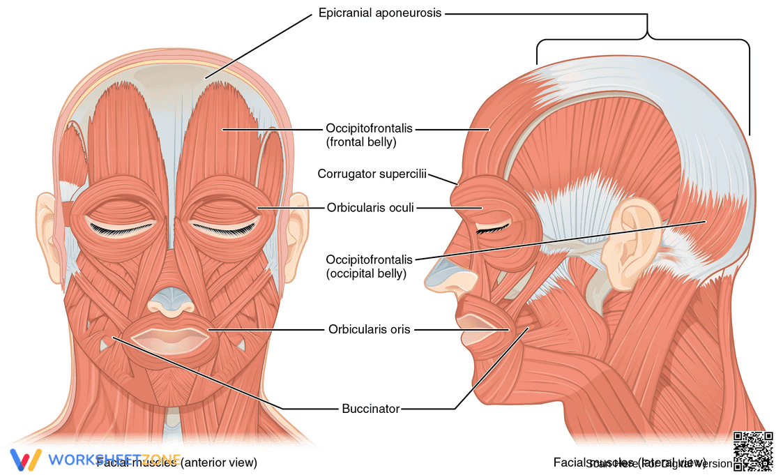
This is an anatomical diagram of the facial muscles. It shows two views: an anterior (front) view and a lateral (side) view. Key muscles are labeled, including the epicranial aponeurosis, occipitofrontalis (frontal and occipital belly), corrugator supercilii, orbicularis oculi, orbicularis oris, and buccinator. The diagram provides a visual representation of the location and structure of these muscles.
Grade Level Suitability:
This worksheet is suitable for high school and college-level anatomy and physiology courses. The detailed labeling and anatomical accuracy require a more advanced understanding of biology. It is also suitable for medical students or those in related healthcare fields.
Why Use It:
This diagram helps students learn and memorize the names and locations of the major facial muscles. It aids in understanding the relationship between muscle structure and facial expressions. It also provides a visual aid for understanding the anterior and lateral views of the muscles.
How to Use It:
Use the diagram to study the location and names of the facial muscles. You can use it for self-testing by covering the labels and identifying the muscles. Compare the anterior and lateral views to understand the 3D arrangement of the muscles. This can be used as a study aid in conjunction with textbooks or online resources.
Target Users:
This worksheet is ideal for students studying anatomy, physiology, biology, or medicine. It is also useful for healthcare professionals, such as nurses and physical therapists, who need a reference for facial muscle anatomy.